<?xml version="1.0" encoding="UTF-8"?><feed xmlns="http://www.w3.org/2005/Atom" xml:lang="en"><title>IndiaBioscience - News from 2020</title><link
    rel="alternate"
    href="https://indiabioscience.org/news/2020"
    type="text/html"
    /><link
    rel="self"
    href="https://indiabioscience.org/news/2020/feed"
    type="application/atom+xml"
    /><id>https://indiabioscience.org/news/2020/feed</id><updated>2026-06-22T02:11:33+05:30</updated><entry><title>A new study explains how the human brain recognizes jumbled words</title><link
                  rel="alternate"
                  href="https://indiabioscience.org/news/2020/a-new-study-explains-how-the-human-brain-recognizes-jumbled-words"
                  type="text/html"
                  /><summary type="html"><![CDATA[
                <p>Our brains are quite proficient at recognizing jumbled words and reading them correctly. Researchers from the Indian Institute of Science, Bengaluru, studied this fascinating phenomenon and came up with a computational model that uses artificial neurons to simulate the way the brain processes jumbled words.</p>              ]]></summary><id>tag:indiabioscience.org,2020-12-30:/news/2020/a-new-study-explains-how-the-human-brain-recognizes-jumbled-words</id><published>2020-12-30T09:00:00+05:30</published><updated>2021-01-05T14:31:31+05:30</updated><author><name>Joel P. Joseph</name><uri>https://indiabioscience.org/authors/rbydKYRYdp1AJjG</uri></author><content type="html"><![CDATA[
                
<p>Our brains are quite proficient at recognizing jumbled words and reading them correctly. Researchers from the Indian Institute of Science, Bengaluru, studied this fascinating phenomenon and came up with a computational model that uses artificial neurons to simulate the way the brain processes jumbled words.</p><figure><a href="https://indiabioscience.org/news/2020/a-new-study-explains-how-the-human-brain-recognizes-jumbled-words"><img
                width="720"
                height="441"
                style="max-width: 100%; height: auto"
                src="https://cdn.indiabioscience.org/media/articles/featured2jumbled.jpg"></a></figure><p>How does our brain read jumbled words correctly? Scientists led by SP Arun and K V S Hari from the Centre for Neuroscience, Indian Institute of Science (IISc), Bengaluru, <a href="https://elifesciences.org/articles/54846">have developed</a> a computational model that sheds light on this. According to this model, when we see a string of letters, our brain uses the letter shapes to form an image of the word and compares it with the closest visually similar word stored in our brain.</p><p>Reading words is a complex process in which our brain decodes the letters and symbols in the word (also called the orthographic code) to derive meaning. Earlier research has shown that our brain processes jumbled words at various levels - visual, phonological and linguistic. </p><p>At the visual level, it is easy to read a jumbled word correctly when the first and the last letters are retained and the other letters are jumbled or replaced with letters of similar shapes. Yet, some arrangements are easier to read than others. For instance, ‘UNIEVRSITY’ is easier to read than ‘UTISERVNIY.’ We can also read words when numbers of similar shape replace letters, e.g. “7EX7__WI7H__NUM83R5.” </p><p>At a linguistic level, it is easier to recognize words that we encounter more frequently or have frequently-used letters. At the phonological level, it is easier to recognize similar sounding words, e.g. tar/car, pun/fun etc. However, how these factors contribute individually or collectively to recognize words remains unclear. </p><p>“We show that our ability to read jumbled words comes from simple rules in the visual system, whereby the response to a string of letters is a weighted sum of its individual letters,” Aakash Agarwal, first author of the paper, says. </p><p>
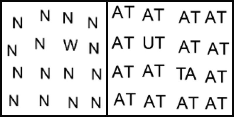
The team asked fluent English speakers aged 22-27 years to search for the odd letter out within a group of letters (distractors) displayed on the screen. The researchers found that the more similar were the shapes of the odd letter and the distractors, the more time the subjects took to accurately spot it. The team could thus calculate an index of how similar or different English letters were to each other, based on the time taken by the subjects to spot the odd letter in this experiment. </p><p>Using this information, the team proceeded to design computational units (artificial neurons) that were mathematically tuned to gauge how similar or different letters were to each other, thereby mimicking the neurons in the brain. Using these artificial neurons, the team then predicted how much time human subjects would take to identify odd two-letter combinations hidden within an array of two-letter distractors and found that the predictions matched the experimental findings.</p><figure><img src="https://cdn.indiabioscience.org/media/articles/collage_2020-12-29-155300.png" data-image="211832"><figcaption>Left: An example of finding the odd letter from a group of distractors. Right: An example of finding the odd two-letter combination (bigram) from a group of distractors (Image: Adapted from Agrawal et al, eLife, 2020)</figcaption></figure><p>The researchers then tested the responses of the artificial neurons to four, five and six-letter words, studying how difficult or easy it was for these neurons to distinguish actual words from jumbled words. The more similar the shape of the jumbled word was to the correct word, the more difficult it was to identify it as jumbled. It was also more difficult to spot jumbled words when the first and the last letters were kept the same. For example, it was easier to spot EPNCIL as a jumbled word than PENICL.</p><p>The artificial neurons processed these words by adding up the responses to individual letters contained within the word. They could also predict the time that the human brain would take to correctly identify a non-jumbled word from within a group of jumbled words. This was confirmed by experimental findings.</p><p>Finally, the researchers used functional MRI to capture brain images of volunteers performing a word recognition task to see which region of the brain got activated during the process. They found that observing a word activates the lateral occipital region – the part of the brain that processes visual information. Following this, the brain compares it with similar-looking words, which is probably stored in the visual word form area (VWFA).</p><figure><grammarly-extension style="position: absolute; top: 0px; left: 0px; pointer-events: none;" class="cGcvT"></grammarly-extension><img src="https://cdn.indiabioscience.org/media/articles/collage2.png" data-image="211833"><figcaption spellcheck="false">Left: The Lateral occipital region is activated upon seeing jumbled words and is computed with the stored words in the VWFA. Right: How our brain reads jumbled words (Image: S P Arun)</figcaption></figure><p><br>“We hope that our model would address the shortcoming of existing models that aim to crack the orthographic code and compel researchers to reconsider the contribution of vision in orthographic processing,” Agarwal says. </p><p>Arpan Banerjee, Additional Professor, National Brain Research Centre (NBRC), who was not involved in the study, says that one unique aspect of this study is how computational modelling has been used to explain the neural data. </p><p>The findings could help in developing efficient text recognition algorithms and also enable better diagnosis of reading disorders like dyslexia. “It would be interesting to see how this model can work for more complex languages like Hindi and Urdu,” Banerjee says. He also wonders if the properties of the artificial neurons would change after learning two languages.</p><p>The team intends to explore the role of visual processing in predicting reading fluency in children. “While deficits in phonological processing are the main cause for dyslexia, a subset of children has deficits at the visual level. We aim to identify this subset through visual experiments and develop training regimes to help improve their reading fluency in our next follow-up study,” Agarwal says. </p>
              ]]></content><category term="neuroscience" label="Neuroscience" /><category term="research" label="Research" /></entry><entry><title>Infosys Prize 2020 winners announced</title><link
                  rel="alternate"
                  href="https://indiabioscience.org/news/2020/infosys-prize-2020-winners-announced"
                  type="text/html"
                  /><summary type="html"><![CDATA[
                <p>In a virtual ceremony on 2 December 2020, the Infosys Science Foundation announced the winners of the twelfth Infosys Prize awards. The awardees represented a diversity of fields and research areas, spread across six categories: Engineering and Computer Science, Humanities, Life Sciences, Mathematical Sciences, Physical Sciences, and Social Sciences</p>              ]]></summary><id>tag:indiabioscience.org,2020-12-08:/news/2020/infosys-prize-2020-winners-announced</id><published>2020-12-08T09:00:00+05:30</published><updated>2020-12-19T19:42:12+05:30</updated><author><name>Susheela</name><uri>https://indiabioscience.org/authors/ANQdMn8qkRK6OE7</uri></author><content type="html"><![CDATA[
                
<p>In a virtual ceremony on 2 December 2020, the Infosys Science Foundation announced the winners of the twelfth Infosys Prize awards. The awardees represented a diversity of fields and research areas, spread across six categories: Engineering and Computer Science, Humanities, Life Sciences, Mathematical Sciences, Physical Sciences, and Social Sciences</p><figure><a href="https://indiabioscience.org/news/2020/infosys-prize-2020-winners-announced"><img
                width="720"
                height="441"
                style="max-width: 100%; height: auto"
                src="https://cdn.indiabioscience.org/media/articles/Pages-from-prize-2020-interactive.jpg"></a></figure><p>The Infosys Science Foundation (ISF), a not-for-profit trust, announced its twelfth Infosys Prize awards on Wednesday, December 2, 2020, on a virtual platform. The prestigious award includes a gold medal, citation, and a cash prize of USD 100000.</p><p>The award recognizes and celebrates outstanding contributions from researchers of Indian origin across six categories: Engineering and Computer Science, Humanities, Life Sciences, Mathematical Sciences, Physical Sciences, and Social Sciences. </p><p>The jury comprised eminent scientists from all over the world represented by six jury chairs: Arvind (Engineering and Computer Science), Kaushik Basu (Social Sciences), Akeel Bilgrami (Humanities), Chandrashekhar Khare (Mathematical Sciences), Shrinivas Kulkarni (Physical Sciences), and Mriganka Sur (Life Sciences).</p><h2><strong>The awardees</strong></h2><p><strong>Hari Balakrishnan</strong></p><p><em>Fujitsu Professor of Computer Science, MIT, USA</em></p><figure style="max-width: 248px; float: left; width: 336px; margin: 0px 20px 20px 0px;"><img src="https://cdn.indiabioscience.org/media/articles/Hari-Balakrishnan_01.jpg" data-image="207915" width="248" height="248" alt="Hari Balakrishnan"><figcaption> Hari Balakrishnan (Photo: ISF)</figcaption></figure><p>The Infosys prize 2020 for <strong>Engineering and Computer science</strong> category was awarded to Hari Balakrishnan for his outstanding contribution to mobile telematics. Balakrishnan's work is recognized for its impactful, practical applications. </p><p>He is currently working on helping people drive better and preventing accidents. The project incorporates algorithms that collect real-time data from sensors embedded in mobile phones to understand how people drive. This data, combined with behavioural science, gives feedback to the driver to drive safely. This technology is currently undergoing trials in India.</p><p>Balakrishnan has several iconic projects to his credit and is a successful entrepreneur. He was the first to develop an accurate indoor navigation system called Cricket. Another noteworthy project is the CarTel - real-time mobile sensing and data accumulation system that equips vehicles with sensors to detect surface conditions of roads. "One of my favourite of Hari's projects is the Pothole Patrol, based on CarTel systems," said Arvind, jury chair, while congratulating him. </p><p><strong>Prachi Deshpande</strong></p><p><em>Professor of History, Centre for Studies in Social Sciences, Kolkata</em></p><figure style="float: right; width: 420px; max-width: 247px; margin: 0px 0px 20px 20px;"><img src="https://cdn.indiabioscience.org/media/articles/Prachi-Deshpande_01.jpg" data-image="207916" alt="Prachi Deshpande" width="247" height="247"><figcaption>Prachi Deshpande (Photo: ISF)</figcaption></figure><p>In the <strong>Humanities</strong> category, this year's winner is the eminent historian Prachi Deshpande. She is well-known for her sophisticated treatment of South Asian historiography (the study of how history is written by historians). Deshpande's research focuses on the sociocultural history of historiography, language, and regional identities. </p><p>Her path-breaking book <em>Creative Pasts: Historical Memory and Identity in Western India</em> (Columbia University Press, 2007) closely examines the emergence of modern history-writing practices in Western India from the Maratha period. Her book has had an immense impact on our understanding of the importance of historical memory in shaping regional identities. </p><p>A prolific writer, Deshpande also has several papers, anthologies, and essays to her credit.</p><p><strong>Rajan Sankaranarayanan</strong></p><p><em>Group Leader, Structural Biology Laboratory, Centre for Cellular and Molecular Biology, Hyderabad</em></p><figure style="float: left; width: 420px; max-width: 262px; margin: 0px 20px 20px 0px;"><img src="https://cdn.indiabioscience.org/media/articles/Prof.-Sankaranarayanan_01.jpg" data-image="207917" alt="Rajan Sankaranarayanan" width="262" height="262"><figcaption>Rajan Sankaranarayanan (Photo: ISF)</figcaption></figure><p>Rajan Sankaranarayanan bagged the Infosys Prize in the <strong>Life Sciences</strong> category. The prize recognizes his seminal contribution in understanding one of the most fundamental mechanisms in biology: the error-free translation of the genetic code that makes proteins. "The structural portraits you have generated “speak a thousand words” and reveal selection mechanisms that are conserved across life," said jury chair Mriganka Sur, congratulating him on his award.</p><p>His work has applications in protein engineering and in the drug design of antibiotics and immunosuppressants.</p><p><strong>Sourav Chatterjee</strong></p><p><em>Professor of Mathematics and Statistics, Stanford University, USA</em></p><figure style="float: right; width: 600px; max-width: 255px; margin: 0px 0px 20px 20px;"><img src="https://cdn.indiabioscience.org/media/articles/Sourav-Chatterjee_01.jpg" data-image="207919" alt="Sourav Chatterjee" width="255" height="255"><figcaption>Sourav Chatterjee (Photo: ISF)</figcaption></figure><p>The winner of the Infosys Prize, 2020 in <strong>Mathematical Sciences</strong> was Sourav Chatterjee, a versatile probabilist and an alumnus of the Indian Statistical Institute. He earned his PhD from Stanford. </p><p>His groundbreaking work in probability and statistical physics find particular mention in areas such as fluctuations in random structures, concentration, and super-concentration inequalities. His contribution plays a critical role in emerging fields such as large deviations for random graphs that occur in computing, social, and business networks.</p><p>Congratulating him, jury chair Chandrashekar Khare said, "You are one of the most powerful problem-solvers in the field of your generation." </p><p><strong>Arindam Ghosh</strong></p><p><em>Professor, Indian Institute of Science, Bengaluru</em></p><figure style="float: left; width: 420px; max-width: 262px; margin: 0px 20px 20px 0px;"><img src="https://cdn.indiabioscience.org/media/articles/Arindam-Ghosh_01.jpg" data-image="207914" alt="Arindam Ghosh" width="262" height="262"><figcaption>Arindam Ghosh (Photo: ISF)</figcaption></figure><p>In <strong>Physical sciences</strong>, the Infosys Priz<strong>e</strong> went to Arindam Ghosh, who has contributed immensely in the field of two-dimensional materials for next-generation electronics. His work involves realizing an atomically thin graphene composite that is highly sensitive in converting optical radiation into electric current. These atomically thin, two-dimensional semiconductors for thermoelectric and optoelectronic devices for new generation electronics will impact quantum technology and sensing.</p><p>"We hope your win will inspire many more young people to take up experimental physics," said jury chair Shrinivas Kulkarni in his congratulatory note.</p><figure style="float: right; width: 417px; max-width: 259px; margin: 0px 0px 20px 20px;"><img src="https://cdn.indiabioscience.org/media/articles/Raj-Chetty02.png" data-image="207918" alt="Raj Chetty" width="259" height="259"><figcaption>Raj Chetty (Photo: ISF)</figcaption></figure><p><strong>Raj Chetty</strong></p><p><em>Professor of Economics, Harvard University</em></p><p>The Infosys Prize 2020 in <strong>Social sciences</strong> was awarded to Raj Chetty, one of the youngest tenured professors in Harvard's history. Chetty completed his PhD at the age of 23 from Harvard and returned there as a faculty.</p><p>Chetty's pioneering research identifies barriers to economic opportunity. It develops solutions to help people escape poverty and gain improved life outcomes. His remarkable work has the ability to gain insights from large datasets with the potential to influence major economic shifts.</p><p><strong>Leveraging education for progress</strong></p><p>In his opening address, Narayana Murthy, ISF president and co-founder of Infosys, emphasized that every country that desires to become a developed nation should prioritize higher education and research. "Good ideas are a result of a high-quality education system and a leading-edge research system," said Murthy.</p><p>He added that good ideas also require a mindset of learning to learn, critical and independent thinking, and proactive problem-solving skills.</p><p>"It is these qualities that the winners of the Infosys prize embody, and the Infosys Prize contributes to this mission by honouring those scientists whose work has the potential to improve our world," said Murthy.</p><p>The ISF has initiated other programs to attract youngsters to a career in research. One of them, The National Lecture Series, requires the Infosys prize winners to talk to university students “about the exciting opportunities a research career offers in India,” said Murthy.</p><p>In his acceptance speech, Raj Chetty said that his childhood exposure to science greatly influenced his career in science. This observation was later reflected in his research on data concerning the children of one million US patent holders. "We found that kids who grow up around scientists are much more likely to become inventors themselves," said Chetty.</p><p>Balakrishnan also recalled his endless curiosity as a child which shaped his career as a scientist, adding that "asking why and how are fundamental to research." </p><p>All the awardees acknowledged the role of their teachers at various institutions, who played an active role in inspiring and mentoring them.</p>
              ]]></content><category term="events" label="Events" /></entry><entry><title>Protecting the unprotected in the Bhagirathi basin</title><link
                  rel="alternate"
                  href="https://indiabioscience.org/news/2020/protecting-the-unprotected-in-the-bhagirathi-basin"
                  type="text/html"
                  /><summary type="html"><![CDATA[
                <p>Protecting India's rich biodiversity has usually taken the form of designating protected areas like national parks and wildlife sanctuaries. Several of these are contiguous with larger landscapes that lack such protection status and that form continuous habitat ranges for many animals. In a recent study, a team of Indian researchers have highlighted the need for more focused conservation strategies in the Bhagirathi basin in Uttarakhand.</p>              ]]></summary><id>tag:indiabioscience.org,2020-10-28:/news/2020/protecting-the-unprotected-in-the-bhagirathi-basin</id><published>2020-10-28T09:00:00+05:30</published><updated>2020-12-19T19:42:26+05:30</updated><author><name>Rupsy Khurana</name><uri>https://indiabioscience.org/authors/AGjYBLmdybMy8VJ</uri></author><content type="html"><![CDATA[
                
<p>Protecting India's rich biodiversity has usually taken the form of designating protected areas like national parks and wildlife sanctuaries. Several of these are contiguous with larger landscapes that lack such protection status and that form continuous habitat ranges for many animals. In a recent study, a team of Indian researchers have highlighted the need for more focused conservation strategies in the Bhagirathi basin in Uttarakhand.</p><figure><a href="https://indiabioscience.org/news/2020/protecting-the-unprotected-in-the-bhagirathi-basin"><img
                width="720"
                height="440"
                style="max-width: 100%; height: auto"
                src="https://cdn.indiabioscience.org/media/articles/BhagirathiFeatured.jpg"></a></figure><p>Delineating protected areas is essential to the conservation of species and landscapes. In a country as populous as India, wildlife and humans often have to scramble for space and resources. Sometimes, wild animals occupy habitats that either exist outside of a protected area or are embedded in the human settlements. It is important to recognize such non-protected areas and implement effective conservation strategies.</p><p>A recent <a href="https://www.cambridge.org/core/journals/oryx/article/mammals-of-the-bhagirathi-basin-western-himalaya-understanding-distribution-along-spatial-gradients-of-habitats-and-disturbances/16FB8B170C0C9ACA921ABFC2F8B25E5E">study</a> conducted by a team of researchers led by Sathyakumar Sambandam, senior scientist at <a href="https://indiabioscience.org/orgs/wii">Wildlife Institute of India, Dehradun</a> highlights one such landscape - the Bhagirathi basin in Uttarakhand - as an important stronghold of biodiversity.</p><p>The Himalayas were carved out millions of years ago by the abrasive energy of flowing rivers and glaciers, aided by the slow accumulation of soil and rocks. The Bhagirathi basin in the Western Himalayas is formed by the Bhagirathi river, a major tributary of the river Ganga. This region provided the researchers with a natural set-up to assess wildlife diversity and its interaction with human activities.</p><p>The basin encompasses diverse habitats and climates ranging from cold and dry with sparse vegetation in the Trans-Himalayas to dense Sal forests in the lower Himalayas. The Gangotri National park is the only protected area in the basin and it largely protects habitats in the high altitudes. In contrast, forests in the lower altitudes are set within patches of human habitations dotted with agricultural fields.</p><p>During the 3-year-long study, researchers placed camera traps at 209 locations spread evenly throughout the Bhagirathi basin. They recorded 39 species of large and medium-sized mammals, including endangered species such as the Bengal tiger, Himalayan brown bear, dhole, and musk deer. The photo captures also revealed the presence of snow leopards, common leopards, Asiatic black bears and Sambar, which are listed as “threatened” by the International Union of Conservation of Nature (IUCN).</p><figure><img src="https://cdn.indiabioscience.org/media/articles/Leopard2.jpg" data-image="202212" alt="Common Leopard in the basin capture in the camera trap"><figcaption>Common Leopard in the basin captured in a camera trap (Photo: WII-DST-NMSHE)</figcaption></figure><p>Interestingly, researchers recorded the presence of Argali, Tibetan sand foxes, woolly hares, Eurasian lynxes and woolly flying squirrels, which had never been sighted in Uttarakhand prior to this survey. This underscores the significance of wildlife habitats and the need for intensive surveys in the Bhagirathi basin.</p><p>The people residing in the basin have traditionally been dependent on the local forests for their household needs. But the burgeoning population, increasing tourism, and pilgrimage activities have further strained the resources of this mountainous region.</p><p>Sathyakumar’s study indicates high rates of human movement in all the habitats during summer, coupled with considerable overlap in human and animal activity. Gangotri National Park witnessed higher activities of feral dogs and livestock during summer as compared to winter. Since feral dogs can transmit diseases and hunt wild prey, their increasing population is another challenge to wildlife management. Images from camera traps placed in subalpine habitats have revealed the presence of hunting activities in the basin.</p><p>Habitat alterations due to development projects can also have grave repercussions on the wildlife in the basin. The recently declared Char Dham Railway Project and the ongoing All-Weather Char Dham Road Project may lead to undesirable changes in some of the pristine habitats in this landscape. According to Ranjana Pal, the first author of the study, while developmental activities are essential to economic growth, an integrated approach acknowledging the importance of wildlife and habitats is imperative for ecosystem health.</p><figure><img src="https://cdn.indiabioscience.org/media/articles/Leopard1.jpg" data-image="202211" alt="Snow Leopard in the basin"><figcaption>Snow Leopard captured in a camera trap (Photo: WII-DST-NMSHE)</figcaption></figure><p>The study concludes that the Bhagirathi Basin is home to a significant amount of biodiversity and needs immediate attention. Since such landscapes lack protection, they are highly fragmented and the wildlife residing there may witness multi-faceted consequences due to intense human activities. Species with large home ranges require movement between different habitats to forage for food and find mates. Corridors through which such movement can take place are also crucial to avoid inbreeding inside the protected areas and hence, maintain genetically healthy populations. High intensity of human activities outside and at the edges of protected areas can lead to loss of connectivity between habitats leading to functional isolation of species inside protected areas. This can, in turn, imperil conservation strategies.</p><p>Apart from losing critical ecological corridors to developmental activities, large mammals, especially carnivores, are threatened due to human-wildlife conflict. In human-dominated landscapes, the risk of human-wildlife conflict is disproportionately high and threatens the continued survival of species.</p><p>“Protected areas cover only a small proportion of India's natural landscapes. Several areas outside protected areas also support populations of wild mammals and help in maintaining connectivity among protected areas,” says Prachi Thatte, coordinator for connectivity conservation, <a href="https://indiabioscience.org/orgs/wwf-india">World Wildlife Fund-India</a>, who was not associated with the study.</p>
              ]]></content><category term="environmental-sciences" label="Environmental Sciences" /><category term="ecology" label="Ecology" /><category term="conservation-biology" label="Conservation Biology" /><category term="research" label="Research" /></entry><entry><title>Scientists discover a new anti-cancer bioactive compound from bacteria</title><link
                  rel="alternate"
                  href="https://indiabioscience.org/news/2020/scientists-discover-a-new-anti-cancer-bioactive-compound-from-bacteria"
                  type="text/html"
                  /><summary type="html"><![CDATA[
                <p>The bacterial world contains a treasure trove of potent compounds with biological activities that can be harnessed for human benefit. Researchers from CSIR-National Chemical Laboratory, Pune and the Jawaharlal Nehru Tropical Botanic Garden and Research Institute, Thiruvananthapuram, have recently found that Urdamycin, a compound produced by <em>Streptomyces </em>bacteria, has the ability to induce cell death in cancer cells.</p>              ]]></summary><id>tag:indiabioscience.org,2020-10-26:/news/2020/scientists-discover-a-new-anti-cancer-bioactive-compound-from-bacteria</id><published>2020-10-26T11:30:00+05:30</published><updated>2020-12-19T19:52:18+05:30</updated><author><name>Susheela</name><uri>https://indiabioscience.org/authors/ANQdMn8qkRK6OE7</uri></author><content type="html"><![CDATA[
                
<p>The bacterial world contains a treasure trove of potent compounds with biological activities that can be harnessed for human benefit. Researchers from CSIR-National Chemical Laboratory, Pune and the Jawaharlal Nehru Tropical Botanic Garden and Research Institute, Thiruvananthapuram, have recently found that Urdamycin, a compound produced by <em>Streptomyces </em>bacteria, has the ability to induce cell death in cancer cells. </p><figure><a href="https://indiabioscience.org/news/2020/scientists-discover-a-new-anti-cancer-bioactive-compound-from-bacteria"><img
                width="720"
                height="440"
                style="max-width: 100%; height: auto"
                src="https://cdn.indiabioscience.org/media/articles/Urdamycin-01.png"></a></figure><p>In a bid to find natural therapeutic routes to cancer treatment, scientists are actively researching novel bioactive chemicals produced by bacteria. Although some such compounds find extensive use in chemotherapy in combination with other drugs, most have limited efficacy and are known to cause side effects.</p><p>Now, a team of scientists led by Syed Dastager from <a href="https://indiabioscience.org/orgs/csir-ncl">CSIR-National Chemical Laboratory, Pune</a>, and Vipin Mohan Dan from the <a href="https://indiabioscience.org/orgs/jawaharlal-nehru-tropical-botanic-garden-and-research-institute-thiruvananthapuram">Jawaharlal Nehru Tropical Botanic Garden and Research Institute, Thiruvananthapuram</a>, have newly <a href="https://pubs.acs.org/doi/10.1021/acschembio.0c00026">discovered the therapeutic potential of a compound called Urdamycin</a> in treating cancer.</p><p>Their study shows that this bioactive bacterial compound has a better scope as an anti-cancer drug compared to Rapamycin - a drug currently widely used in combination in chemotherapy. The study reveals that Urdamycin not only binds to a critical cell growth protein but also induces tumour cell death in two different ways.</p><p>Bacteria live under constant hostility from various environmental factors, including competition from other microbes. To thrive, many of them have developed toxins to ward off invaders, or chemical messengers to carry out social communication within their community. Some higher-order bacteria living in regions of rich biodiversity produce compounds that can have clinical use as drugs. Hence, such bacteria are extensively exploited, one example being the <a href="https://www.sciencedirect.com/science/article/pii/S1413867012001341#:~:text=Streptomyces%20is%20a%20genus%20of,into%20a%20chain%20of%20spores."><em>Streptomyces</em></a> group which produces many bioactive compounds, including one called Rapamycin.</p><p>Initially, Rapamycin drew attention as an anti-cancer drug (often used in combination with other chemotherapeutic medications), though its exact mechanism of action remained unknown for many years. About two decades ago, scientists revealed that Rapamycin bound to a critical growth protein called <a href="https://jcs.biologists.org/content/122/20/3589">mTOR</a> (mechanistic or mammalian Target of Rapamycin), and thus inhibited tumour cell growth.</p><p>Further investigations revealed that mTOR forms a complex of proteins, including two forms - mTORC1 and mTORC2, both of which play important roles in cell growth. However, Rapamycin only binds mTORC1, thereby <a href="https://www.hindawi.com/journals/omcl/2017/1726078/">limiting its</a> ability to induce cancer cell death. Hence, the requirement for more efficient mTOR inhibitors re-emerged and became a significant area of interest.</p><p>"In an attempt to discover novel bioactive bacterial compounds, we explored the rich bacterial biodiversity of the Western Ghats," says Dan. He was instrumental in identifying the bacterial species <em>Streptomyces</em> OA293 that produces Urdamycin.</p><p>The process of identifying, extracting and analysing suitable compounds for cancer treatment is an arduous one. Also, it is challenging to synthesise and purify them from bacterial cultures in the laboratory, or to ascertain their molecular structures.</p><p>To overcome these difficulties, the team employed a multi-pronged strategy. First, they screened the genetic material of the Streptomyces OA293 by <a href="https://en.wikipedia.org/wiki/Whole_genome_sequencing">whole-genome analysis</a>. This allowed them to list gene clusters that might be able to produce potential anti-cancer compounds. Then they extracted the pure form of the compounds from the bacteria. The extractions were subjected to rigorous biochemical analysis using <a href="https://www.sciencedirect.com/science/article/pii/B9780120885619500065">Nuclear Magnetic Resonance and Mass Spectrometry</a>, which helped the researchers ascertain the molecular structure of the compounds. Finally, the compounds that showed the greatest potential to inhibit mTOR — Urdamycin V and E — were purified and tested on lab cultures of breast and cervical cancer cells.</p><figure><img src="https://cdn.indiabioscience.org/media/articles/UrdamycinWorkflow.JPG" data-image="200763" alt="Urdamycin Workflow"><figcaption>The workflow used by the researchers to isolate and test Urdamycin's anti-cancer activity (Image: Vinodh J S)</figcaption></figure><p>Vinodh J S, a team member, says, "Our research reveals that Urdamycin is a more potent mTOR inhibitor than Rapamycin. It completely inactivates both the components mTORC1 and mTORC2." The study also indicates the possibility that the intracellular binding sites of Urdamycin and Rapamycin are different. Moreover, Urdamycin induces programmed cancer cell death in two ways: by apoptosis (triggering the killing of tumour cells) and by autophagy (triggering starvation of the tumour cells).</p><p>"Our study reveals that Urdamycin completely inhibits Akt activation - another signalling protein, known to be involved in tumour progression and cell survival - leading to cancer cell death," Dastager points out.</p><p>"The biochemical approach employed in this study is solid and well-established. The novel method aided in revealing the exact molecular structure of Urdamycin and the mechanism by which it induces cancer cell death," says Suchetan Pal, assistant professor at the <a href="https://indiabioscience.org/orgs/iit-bhilai">Indian Institute of Technology, Bhilai</a>. He was not involved in the study.</p><p>Collectively, the initial results indicate that Urdamycin has potential therapeutic value for cancer treatment and can be scaled up for further analysis, suggests Pal.</p>
              ]]></content><category term="biochemistry" label="Biochemistry" /><category term="molecular-biology" label="Molecular Biology" /><category term="research" label="Research" /></entry><entry><title>Shall we grow in the dark? A COP1 story</title><link
                  rel="alternate"
                  href="https://indiabioscience.org/news/2020/shall-we-grow-in-the-dark-a-cop-story"
                  type="text/html"
                  /><summary type="html"><![CDATA[
                <p>Plants react to their immediate environment in a number of ways and use the information so gleaned to make crucial decisions about growth and survival. In a new study, researchers from the Indian Institute of Science Education and Research (IISER) Bhopal have discovered a new molecular mechanism via which seedlings react to the absence of light and use it to modulate their growth rate to ensure optimum conditions for survival.</p>              ]]></summary><id>tag:indiabioscience.org,2020-10-09:/news/2020/shall-we-grow-in-the-dark-a-cop-story</id><published>2020-10-09T09:00:00+05:30</published><updated>2020-12-19T19:52:05+05:30</updated><author><name>Divya Sriram</name><uri>https://indiabioscience.org/authors/DivyaSriram</uri></author><content type="html"><![CDATA[
                
<p>Plants react to their immediate environment in a number of ways and use the information so gleaned to make crucial decisions about growth and survival. In a new study, researchers from the Indian Institute of Science Education and Research (IISER) Bhopal have discovered a new molecular mechanism via which seedlings react to the absence of light and use it to modulate their growth rate to ensure optimum conditions for survival. </p><figure><a href="https://indiabioscience.org/news/2020/shall-we-grow-in-the-dark-a-cop-story"><img
                width="720"
                height="440"
                style="max-width: 100%; height: auto"
                src="https://cdn.indiabioscience.org/media/articles/plant-3117525_960_720.jpg"></a></figure><p>Seeds germinate only when they find the right environmental conditions. Once germinated, the seedling starts developing in preparation for emerging above the ground and commencing life as an independent entity. But what happens when the conditions are not optimum <em>after</em> the seed has already germinated? One way that this can happen is if the seedling gets buried under the soil, resulting in prolonged dark conditions.</p><p><a href="https://onlinelibrary.wiley.com/doi/full/10.1111/tpj.14844">In a recent study</a>, Sourav Datta, Associate Professor, <a href="https://indiabioscience.org/orgs/iiser-bhopal">Indian Institute of Science Education and Research (IISER), Bhopal</a> and his group show that just as a seed does not germinate if the conditions are not right, seedlings also do not grow unless certain conditions are met. In particular, the authors studied the importance of the presence of light for the growth and survival of seedlings.</p><p>The researchers uncovered a mechanism by which seedlings can perceive when it is dark, resulting in a pause or arrested growth in the absence of sufficient light. This involves a plant growth hormone called abscisic acid (ABA).</p><p>Plant cells receive signals from the environment (light, temperature, pH, humidity), through sensors present on their surface. They also secrete plant hormones like ABA, auxins, gibberellins, etc. that serve a variety of functions. Signals from the sensors as well as the plant hormones instruct the cells to carry out the appropriate metabolic activities required for a given stage of plant growth. </p><p>This is facilitated through a series of signals passed on from one molecule to another, like a baton in a relay race. Every environmental cue results in activation of a specific set of signalling pathways, helping the plant make complex decisions. Identifying the molecular players in this relay race is vital to studying plant functions and growth. </p><p>When light is optimum for seedling growth, a certain signalling pathway is activated through light-sensing photoreceptors on the cell surface, providing a “go” signal for growth and development. Datta’s group found that in darkness (caused by the seedling getting buried under soil layers), a molecule called COP1 communicates with the plant hormone ABA, arresting the growth of developing seedlings. </p><figure><img src="https://cdn.indiabioscience.org/media/articles/Sourav-dutta.jpg" data-image="199515"><figcaption>The team that worked on this study. From left to right, Sourav Dattta, Yadukrishnan P, Nevedha Ravindran and Rahul PV (Image: Datta lab)</figcaption></figure><p>Datta explains that while seedlings normally slow down their growth in dark conditions, the presence of excess ABA makes seedlings more sensitive to growth arrest. This observation prompted them to hypothesize that there might be a signalling molecule activated only in the dark, working hand-in-hand with ABA to promote growth-arrest. </p><p>Their search pointed towards COP1, a protein found to be active in dark conditions, while less active in the presence of light. The researchers found that when COP1 levels are high in seedlings, they are more responsive to ABA-mediated arrest in seedling development, compared to when it is absent. Using mutant plant lines, they found that seedlings lacking COP1 did not lower their growth rate even in the presence of ABA. These studies suggested that COP1 was the primary player responsible for making seedlings sensitive to ABA-mediated growth arrest.</p><p> Early stages of plant growth are highly regulated and crucial for the survival of seedlings. Newly germinated seedlings face stresses like low light, water scarcity, abnormal pH or salinity of soil, etc, all of which compromise their ability to make food. With limited energy stored in their tiny leaves, they have to conserve energy to adapt and survive in stressful conditions. Thus, prioritizing survival over growth, the seedlings choose to remain in an arrested state under unfavourable conditions, while keeping defence mechanisms active. </p><p>The researchers used the plant model <em>Arabidopsis thaliana </em>for their experiments, but this molecular interplay could be investigated for similar roles in other plants, as COP1 and many components of ABA-associated signalling are conserved across plant species.</p><p>Jitendra K. Thakur, Scientist at <a href="https://indiabioscience.org/orgs/nipgr">National Institute of Plant Genome Research (NIPGR), New Delhi</a>, who was not associated with this study, adds “This study may have implications in agriculture as the optimum timing of seed germination and seedling establishment is critical to ensure high plant yields, which can be utilized to enhance crop productivity especially under changing climatic conditions.”</p>
              ]]></content><category term="plant-biology" label="Plant Biology" /><category term="research" label="Research" /></entry><entry><title>National Bio Entrepreneurship Competition (NBEC) 2020 invites applications</title><link
                  rel="alternate"
                  href="https://indiabioscience.org/news/2020/national-bio-entrepreneurship-competition-nbec-2020-invites-applications"
                  type="text/html"
                  /><summary type="html"><![CDATA[
                <p>A positive highlight of the COVID-19 situation has been the large number of grassroots-level <a href="https://indiabioscience.org/columns/indian-scenario/innovations-to-make-india-self-reliant-in-tackling-covid-19">innovations brought forth</a> by Indian start-ups, entrepreneurs, and researchers to counter the pandemic. A thriving culture of innovation and entrepreneurship is necessary to nurture this spirit. In this regard, the fourth edition of the National Bio Entrepreneurship Competition (NBEC) is currently open for applications.</p>              ]]></summary><id>tag:indiabioscience.org,2020-10-05:/news/2020/national-bio-entrepreneurship-competition-nbec-2020-invites-applications</id><published>2020-10-05T11:26:00+05:30</published><updated>2020-12-19T19:51:55+05:30</updated><author><name>Joel P. Joseph</name><uri>https://indiabioscience.org/authors/rbydKYRYdp1AJjG</uri></author><content type="html"><![CDATA[
                
<p>A positive highlight of the COVID-19 situation has been the large number of grassroots-level <a href="https://indiabioscience.org/columns/indian-scenario/innovations-to-make-india-self-reliant-in-tackling-covid-19">innovations brought forth</a> by Indian start-ups, entrepreneurs, and researchers to counter the pandemic. A thriving culture of innovation and entrepreneurship is necessary to nurture this spirit. In this regard, the fourth edition of the National Bio Entrepreneurship Competition (NBEC), a one-of-a-kind launch platform for bio entrepreneurs in India, is currently open for applications. </p><figure><a href="https://indiabioscience.org/news/2020/national-bio-entrepreneurship-competition-nbec-2020-invites-applications"><img
                width="720"
                height="440"
                style="max-width: 100%; height: auto"
                src="https://cdn.indiabioscience.org/media/articles/NBEC-2020-Featured2.jpg"></a></figure><p>The <a href="https://nationalbioentrepreneurship.in/">National Bio Entrepreneurship Competition (NBEC)</a>, a contest that identifies and nurtures bio-entrepreneurs in India, is organized every year by the <a href="https://indiabioscience.org/orgs/c-camp">Centre for Cellular and Molecular Platforms (C-CAMP)</a>. The fourth edition of this competition is currently inviting business ideas in life sciences, covering agriculture and allied fields, healthcare, antimicrobial resistance, digital health, medical devices, environment, industrial biotechnology, and personal care products. The last date to apply is October 7, 2020. </p><p>NBEC is a joint initiative of C-CAMP with <a href="https://indiabioscience.org/orgs/birac">Biotechnology Industry Research Assistance Council (BIRAC)</a> and is supported by <a href="https://nationalbioentrepreneurship.in/events/nbec2020?id=partners">industry partners</a>. It aims to attract and support bio-entrepreneurs in the country with novel, scalable, deep-tech business ideas that have social impact. “NBEC has become a national platform to amplify the spirit of bio-entrepreneurship across India, with young and exciting innovative ideas being assessed and supported through NBEC and further with industry and investors,” says Taslimarif Saiyed, CEO and Director, C-CAMP. </p><p>The competition has two application tracks: (i) student teams, (ii) start-ups and individuals. A student team must have four members who are Indian citizens currently pursuing a graduate, postgraduate, or PhD degree. If the students have formed a company, the company must be less than two years old at the time of applying.</p><p>In the ‘start-ups and individuals’ category, start-ups, Micro, Small, and Medium Enterprises (MSMEs), large companies, post-doctoral researchers, faculty, scientists, working professionals, and aspiring entrepreneurs can apply. The applicants must be citizens of India. Companies that apply must be registered in India and have a majority Indian ownership. </p><p>The competition, held online this year, has four rounds which will be spread over the next four months. In round one, an expert panel consisting of members from the industry, academia, and investor community will screen the proposals. In the second round, the selected applicants will present their idea to a jury panel that will choose the best few business ideas. In round three, the selected candidates will attend an entrepreneurship development boot camp for two days, following which they will present their business plan before a jury panel. In the fourth and final round, the finalists will pitch their ideas before a grand jury, who will select the winners based on the scientific validity of the idea, feasibility of the proposed solution and business, and novelty of the approach.</p><p>For the winning student teams, the competition offers total cash prizes of up to Rs 10 lakhs. The winners in the ‘start-ups and individuals’ category will take away total cash prizes of Rs 7.1 crores, as well as investment opportunities.</p><p>“A unique feature of NBEC is that it doesn’t just end the day when winners are announced; it then serves as a launchpad for the competitors with the best ideas,” write Nisha Holla, Technology Fellow, and Taslimarif Saiyed, CEO and Director, C-CAMP, in an article <a href="https://www.financialexpress.com/opinion/science-based-solutions-india-needs-more-platforms-like-national-bio-entrepreneurship-competition/1864975/">published</a> in <em>Financial Express </em>in February, 2020.</p><p>The winners of the 2019 edition of NBEC included <a href="https://medium.com/@iamdiv97">Divanshu Kumar</a> of Alcheme Robotics, Mayur Shetty of <a href="https://www.blackfrog.in/">Blackfrog Technologies</a>, Ragul Paramasivam of <a href="https://www.chimertech.com/">Chimertech Innovations LLP</a>, Shomeshwar Singh of <a href="https://seesoundlive.com/">4S Medical Research</a>, Prakhar Jain of <a href="https://microxlabs.com/">MicroX Labs</a>, Rajesh Nandipati of <a href="https://oncosimis.com/">Oncosimis Biotech</a>, Asawari Kane of <a href="https://padcarelabs.com/">PadCare Labs</a>, Arindam Ghatak of <a href="https://www.biomoneta.com/">Biomoneta</a> and Halagappa Eswarappa Shashidhar of <a href="http://cultiva.in/">Cultiva AgriTech</a>. </p><p>Some of the winning ideas were robotic solutions to clean septic tanks, portable battery-powered refrigerators for last-mile vaccine delivery, smokeless sanitary napkin disposal units, assistive technology for hearing-impaired individuals that converts their speech effort into a visual pattern, and solutions for sugarcane cultivation. </p><p>Beyond the awards, NBEC paves the way for mentorship and networking with industry leaders. Partners like Biocon, Kotak, Novozymes and Ankur seeds have continued to mentor some of the winners after the competition. </p><p>“This year, we have added new categories to specifically encourage young students’ innovative ideas and new start-ups. These categories would be in addition to the main category which is open to all other start-ups and applicants. We hope that this will further widen this national platform’s impact across all India,” says Saiyed.</p><p>“When I applied for NBEC 2019, I had barely any idea about entrepreneurship in life sciences. I had only applied to it thinking it’s an idea competition and that there would be prizes. But the journey with NBEC 2019 so far has been so enriching in terms of discussions with peers and mentors here that it really moulded me into taking up entrepreneurship,” says Aparna Nair, a finalist in NBEC2019.<br></p>
              ]]></content><category term="biotechnology" label="Biotechnology" /><category term="entrepreneurship" label="Entrepreneurship" /><category term="events" label="Events" /></entry><entry><title>Monsoon Brain Meeting 2020: Talking about the brain in the rain</title><link
                  rel="alternate"
                  href="https://indiabioscience.org/news/2020/monsoon-brain-meeting-2020-talking-about-the-brain-in-the-rain"
                  type="text/html"
                  /><summary type="html"><![CDATA[
                <p>The COVID-19 pandemic has expanded our need for virtual meeting platforms and pushed conference organisers to innovate to ensure that scientific networking continues to flourish in these troubled times. The Monsoon Brain Meeting 2020, held in June 2020, aimed to embrace the virtual format and use it to facilitate discussions on and around neuroscience, as reported here by Annapoorna, one of the attendees of the meeting.</p>              ]]></summary><id>tag:indiabioscience.org,2020-09-21:/news/2020/monsoon-brain-meeting-2020-talking-about-the-brain-in-the-rain</id><published>2020-09-21T09:00:00+05:30</published><updated>2020-12-19T19:51:41+05:30</updated><author><name>Annapoorna P K</name><uri>https://indiabioscience.org/authors/76ldNMzk3kL29rX</uri></author><content type="html"><![CDATA[
                
<p>The COVID-19 pandemic has expanded our need for virtual meeting platforms and pushed conference organisers to innovate to ensure that scientific networking continues to flourish in these troubled times. The Monsoon Brain Meeting 2020, held in June 2020, aimed to embrace the virtual format and use it to facilitate discussions on and around neuroscience, as reported here by Annapoorna, one of the attendees of the meeting. </p><figure><a href="https://indiabioscience.org/news/2020/monsoon-brain-meeting-2020-talking-about-the-brain-in-the-rain"><img
                width="720"
                height="440"
                style="max-width: 100%; height: auto"
                src="https://cdn.indiabioscience.org/media/articles/MonsoonBrainFeatured2.jpg"></a></figure><p>The <a href="http://monsoonbrain.org/">Monsoon Brain Meeting (MBM) 2020</a>, an exclusively virtual neuroscience meeting, was held from 24 to 26 June 2020. It was organised by Arjun Ramakrishnan, <a href="https://indiabioscience.org/orgs/iitk">Indian Institute of Technology (IIT) Kanpur</a> and Venkatakrishnan Ramaswamy, <a href="https://indiabioscience.org/orgs/bits-pilani-hyderabad-campus">Birla Institute of Technology and Science (BITS) Pilani, Hyderabad</a> and funded by <a href="https://indiabioscience.org/orgs/the-wellcome-trust-dbt-india-alliance">DBT/Wellcome Trust India Alliance</a> and Pratiksha Trust. The conference included 16 talks by invited speakers from India and abroad and 140 short talks, covering broad topics in neuroscience. In a special session called ‘Career Spotlights’, 15 postdoctoral researchers presented their work in order to be considered for faculty positions in India. </p><p>In addition to these, there were four panel discussions. A panel called <strong>‘An India Brain Project?’ </strong>was moderated by Upinder Bhalla, <a href="https://indiabioscience.org/orgs/ncbs">National Centre for Biological Science (NCBS), Bengaluru</a>. The panellists discussed the possibility of a major collaboration to utilize the varied expertise of neuroscientists across the nation to better understand different aspects of brain function and dysfunction. The panel laid special emphasis on having regular meetings and presentations with greater involvement of students and young researchers to promote collaborations. Another panel, <strong>‘Neuroscience Careers’,</strong> moderated by Ramakrishnan and including panellists from various scientific professions, discussed some of the career paths that a neuroscientist can embark on. Apart from a research career, the panellists provided perspectives on other less traversed paths, including technology, management, education and advisory roles. </p><p>The other two panels dealt with topics of societal relevance. One of them, <strong>‘Women and Trans persons in Neuroscience’</strong>, was moderated by Vidita Vaidya, <a href="https://indiabioscience.org/orgs/tifr">Tata Institute of Fundamental Research, Mumbai</a>, and had panellists representing different age groups and gender identities. The panellists discussed how most people with a gender identity other than cisgender male face some kind of discrimination or prejudice. The panel members shared their experiences and highlighted the fact that the onus is on each of us to call out or raise our voices against discrimination in our circles and places of work. </p><p>A panel called <strong>‘Science for All and All for Science’</strong>, hosted by Bittu K Rajaraman, <a href="https://indiabioscience.org/orgs/ashoka-university">Ashoka University, Sonipat</a>, discussed better inclusion and support of diverse groups in neuroscience irrespective of gender, ethnicity, economic or social background. The panellists included Angela Saini, Science Journalist and Author; Abha Sur, Scientist, Historian, Author and Lecturer in Massachusetts Institute of Technology; and Sonajharia Minz, Vice-Chancellor, <a href="https://indiabioscience.org/orgs/sido-kanhu-murmu-university">Sido Kanhu Murmu University, Jharkhand</a>. The panellists talked about their respective initiatives in raising awareness and fighting discrimination against minority groups. They agreed that going ahead, Indian academia needs a radical change in thought and system to be more inclusive and supportive. </p><p>“Panels that address such topics are very important and Indian academia needs them, which made me really happy to be a part of the organising team,” said Aastha Sharma, one of the student organisers. </p><p>The final session of the meet was a talk titled <strong>‘Growing up in Science’</strong> by K VijayRaghavan, Principal Scientific Advisor, Government of India, in which he spoke about his journey leading up to a career in science. There were also a couple of contests for the artists and writers in the neuroscience community. The winner of the <a href="http://monsoonbrain.org/con1.html">NeuroArt contest</a> was Karthik Krishnamurthy, Thomas Jefferson University, USA. The <a href="http://monsoonbrain.org/con2.html">NeuroFiction contest</a> had two winners, Samatha Mathew, <a href="https://indiabioscience.org/orgs/csir-igib">CSIR-Institute of Genomics and Integrative Biology</a>, Delhi and Sidra Yaqoob, Duke-National University of Singapore Medical School. </p><p>The entire conference was open for all with no registration fee and had almost 800 participants, several of whom are young researchers and undergraduate students. Despite being online, due to the user-friendly interface of Crowdcast, there was immense interaction among the attendees over the chatbox. The hosts also ensured that the participants engaged with the speakers. The participants could ask questions by typing into a ‘Questions’ tab on-screen and the moderators invited some of them over video so that they could interact with the speakers face to face. </p><p>“I was overjoyed by the turnout, participation, high quality of talks and panels, and the fun the attendees had interacting with one another and the speakers. Overall a win-win for neuroscience and the community!” said Ramakrishnan.</p><p>Several people contributed towards making the meeting a success. As Ramaswamy said, “We received a great deal of generous help from everyone we asked, the funding agencies, the invited speakers and panellists and all the student volunteers.” The hope is that MBM will set an example for more such meets in the future, which will keep science and enthusiasm alive even in tough times. “Many have volunteered to help us organise such events because they are greedy for more. It would be great to enable more such meetings,” said Ramakrishnan. <br></p>
              ]]></content><category term="neuroscience" label="Neuroscience" /><category term="networking" label="Networking and Collaboration" /><category term="events" label="Events" /></entry><entry><title>A new study explains how the tuberculosis bacteria activates HIV</title><link
                  rel="alternate"
                  href="https://indiabioscience.org/news/2020/a-new-study-explains-how-the-tuberculosis-bacteria-activates-hiv"
                  type="text/html"
                  /><summary type="html"><![CDATA[
                <p>Tuberculosis is a common comorbidity in those infected with HIV/AIDS, and the two conditions are known to exacerbate one another. A new study from researchers at the Indian Institute of Science, Bengaluru demonstrates that <em>Mycobacterium tuberculosis, </em>the tuberculosis bacteria, can reactivate dormant HIV within the human body, a process that can potentially be targeted by specific drugs<em>. </em></p>              ]]></summary><id>tag:indiabioscience.org,2020-09-14:/news/2020/a-new-study-explains-how-the-tuberculosis-bacteria-activates-hiv</id><published>2020-09-14T09:00:00+05:30</published><updated>2020-10-14T10:39:58+05:30</updated><author><name>Joel P. Joseph</name><uri>https://indiabioscience.org/authors/rbydKYRYdp1AJjG</uri></author><content type="html"><![CDATA[
                
<p>Tuberculosis is a common comorbidity in those infected with HIV/AIDS, and the two conditions are known to exacerbate one another. A new study from researchers at the Indian Institute of Science, Bengaluru demonstrates that <em>Mycobacterium tuberculosis, </em>the tuberculosis bacteria, can reactivate dormant HIV within the human body, a process that can potentially be targeted by specific drugs<em>. </em></p><figure><a href="https://indiabioscience.org/news/2020/a-new-study-explains-how-the-tuberculosis-bacteria-activates-hiv"><img
                width="720"
                height="440"
                style="max-width: 100%; height: auto"
                src="https://cdn.indiabioscience.org/media/articles/HIV-budding-Color-resized.jpg"></a></figure><p>Taken together, tuberculosis (TB) and Human Immunodeficiency Virus/Acquired Immunodeficiency Syndrome (HIV/AIDS) cause <a href="https://www.who.int/news-room/fact-sheets/detail/the-top-10-causes-of-death">over 2.2 million deaths</a> each year worldwide. The two also often occur simultaneously, with people with HIV being <a href="https://www.who.int/en/news-room/fact-sheets/detail/tuberculosis">more likely to develop TB</a> than people who do not have HIV. <em>Mycobacterium tuberculosis </em>(<em>Mtb</em>), the bacterium that causes TB, is known to enhance the multiplication of HIV. This synergy between the two pathogens hampers the treatment of both the conditions. </p><p>Now, a group of researchers led by Amit Singh, Associate Professor, Centre for Infectious Diseases Research, <a href="https://indiabioscience.org/orgs/iisc">Indian Institute of Science (IISc), Bangalore</a>, <a href="https://mbio.asm.org/content/11/2/e03293-19">has deciphered</a> one of the ways via which TB accelerates AIDS - reactivating dormant HIV within immune cells.</p><p>HIV is classified into two major types: HIV-1 and HIV-2. HIV-1 is the more infectious and predominant of the two. After entering a human, the virus hijacks different types of immune cells – macrophages, lymphocytes, and dendritic cells – and uses them to make copies of itself. It can also remain in an inactive state for many years within some immune cells. <em>Mtb</em>, on the other hand, primarily infects macrophages. </p><p>“HIV and <em>Mtb</em> can exacerbate each other; however, there are very limited studies that show empirical data for the same,” says Dhiraj Kumar, Group Leader, Cellular Immunology, <a href="https://indiabioscience.org/orgs/icgeb-delhi">International Centre for Genetic Engineering and Biotechnology (ICGEB)</a>, who was not involved in the study. The current study is the first to show that <em>Mtb </em>reactivates HIV-1.</p><p>Singh’s team did their experiments on immune cells cultured in the laboratory. They infected macrophages with <em>Mtb </em>and grew them together with other immune cells infected with HIV-1. In these latter cells, the virus usually remains in an inactive state until it is activated by certain signals. However, the presence of <em>Mtb</em>-infected cells nearby was sufficient to activate the virus. </p><p>The researchers found that <em>Mtb</em>-infected macrophages secreted membrane-bound spherical structures, called <strong>exosomes</strong>, which could, by themselves, activate the virus in the nearby cells. This discovery of the exosomes from <em>Mtb-</em>infected macrophages and their ability to talk to cells that lie far away was the turning point of the study. This could explain how <em>Mtb</em> exacerbated HIV and provided a lead to investigate the molecular pathways involved. </p><p>On probing further, the team found that the exosomes contained several proteins derived from the <em>Mtb</em>-infected macrophages. The researchers mapped these constituents to the molecular pathways they were involved in, identifying two specific pathways (galectins and heat shock proteins) that trigger HIV-1 reactivation. Blocking one of the proteins involved in these pathways with an inhibitor drug could stop the exosomes from reactivating HIV-1. This suggests that this inhibitor might be able to provide a functional treatment for HIV-TB co-infected patients and might improve their quality of life. However, this hypothesis must be tested with more experiments. </p><p>“It is exciting to know that exosomes from <em>Mtb</em>-infected macrophages also package specific molecular factors, which contribute to reactivation of HIV in certain target cells,” Kumar says. “This new mechanism can be exploited to develop novel tools for handling the challenge of co-morbidity associated with HIV.” </p><p>The exosomes also decrease the level of Glutathione, a molecule that protects cells from oxidative stress, and has been linked to HIV-reactivation. This might be another pathway via which the two pathogens synergise. “Interestingly, Glutathione levels are known to be depleted in TB patients and also in the lungs of animals infected with <em>Mtb</em>,” says Singh.</p><p>Singh explains that while this report provides some initial clues of how <em>Mtb </em>reactivates HIV-1 and further work is needed to reveal these mechanisms inside a living system. </p><p>“The significance of our research is in identifying the role of fundamental mechanisms that catalyze HIV-<em>Mtb</em> synergy,” Singh says. “This will greatly enhance our understanding of co-infection [of HIV-1 and <em>Mtb</em>], lead to a wider impact on the biomedical research community and create new translational opportunities.”</p><p>Ramandeep Singh, Associate Professor, <a href="https://indiabioscience.org/orgs/thsti">Translational Health Science and Technology Institute (THSTI)</a>, who was not involved in the study, agrees. “The results have huge translational implications and the study has identified various metabolic/regulatory pathways that can be targeted for design of better therapeutic interventions,” he says. </p>
              ]]></content><category term="health-and-medicine" label="Health &amp; Medicine" /><category term="microbiology" label="Microbiology" /><category term="cell-biology" label="Cell Biology" /><category term="research" label="Research" /></entry><entry><title>Bug story: Looking at Indian Bison domestication through the lens of gut microbes</title><link
                  rel="alternate"
                  href="https://indiabioscience.org/news/2020/bug-story-looking-at-indian-bison-domestication-through-the-lens-of-gut-microbes"
                  type="text/html"
                  /><summary type="html"><![CDATA[
                <p>The gut microbiome of wild animals can provide a plethora of information related to animal health. However, studies looking at evolutionary and animal health-related issues through the lens of gut microbes are currently lacking in India. A recent study reveals the gut bacterial diversity of Indian <em>Gaur</em> and its domesticated form <em>Mithun</em>.</p>              ]]></summary><id>tag:indiabioscience.org,2020-09-07:/news/2020/bug-story-looking-at-indian-bison-domestication-through-the-lens-of-gut-microbes</id><published>2020-09-07T14:00:00+05:30</published><updated>2020-10-14T10:40:24+05:30</updated><author><name>Hina Malik</name><uri>https://indiabioscience.org/authors/GXpwL2eYXBMrb2J</uri></author><content type="html"><![CDATA[
                
<p>The gut microbiome of wild animals can provide a plethora of information related to animal health. However, studies looking at evolutionary and animal health-related issues through the lens of gut microbes are currently lacking in India. A recent study reveals the gut bacterial diversity of Indian <em>Gaur</em> and its domesticated form <em>Mithun</em>.</p><figure><a href="https://indiabioscience.org/news/2020/bug-story-looking-at-indian-bison-domestication-through-the-lens-of-gut-microbes"><img
                width="720"
                height="440"
                style="max-width: 100%; height: auto"
                src="https://cdn.indiabioscience.org/media/articles/Gaur_And_Calf_85173055.jpeg"></a></figure><p>Gut-living bacteria, also known as the gut microbiome, have been shown to play an important role in host health by supporting the immune system, nutrition, development and even influencing behaviour. Understanding the gut microbiome of wild endangered animals and the role it plays in their health could have significant applications in species conservation and management. </p><p>Nevertheless, little research has been done in this area and many of the ways in which the host’s environment and genetics shape the diversity of the gut microbiome remain unknown. Recently, researchers from the <a href="https://indiabioscience.org/orgs/central-university-of-kerala">Central University of Kerala, Kasaragod</a>, in collaboration with the Institute of Infectious Diseases, Switzerland, <a href="https://www.frontiersin.org/articles/10.3389/fmicb.2020.00133/full">published the gut microbiome</a> of the Indian Bison, locally known as "Gaur". The researchers also investigated how domestication influences the gut microbiome, especially in the context of Indian wild animals. </p><p>Gaurs are endemic to South and Southeast Asia and the majority of their populations live in India. They are considered a vulnerable species by IUCN. Gaurs are mainly restricted to national parks in the Western Ghats, central Indian highlands, and Northeastern Himalayas. The domesticated cousin of gaur is "Mithun", which is believed to have diverged from wild gaur more than 8000 years ago. </p><p>For this study, researchers collected faecal samples from both wild and captive gaurs as well as from mithuns. The faecal microbiome, which represents an amalgam of bacteria from different regions of the gut, was analysed by high-throughput sequencing of the 16S rRNA gene of the bacteria, a common approach for such purposes. Interestingly, this analysis revealed that domestication leads to a severe reduction in gut microbial diversity in both captive and domestic forms of gaur in comparison to their wild counterparts.</p><figure><img src="https://cdn.indiabioscience.org/media/articles/Gayal_Mithun.jpg" data-image="189568" alt="Mithun"><figcaption>Domesticated mithun near Darjeeling Para in Bandarban, Bangladesh (Photo: Aditya Kabir / Public domain)</figcaption></figure><p>The study found that two beneficial bacterial families, <em>Ruminococcaceae</em> and <em>Lachnospiraceae</em>, which play vital roles in digestion in herbivores, showed lower abundance in captive and domestic populations. Both bacterial families are also important members of the human microbiome. The bacterial composition also suggested that captive and domestic gaur had impaired metabolism and immune function compared to wild gaur. According to Wasimuddin, a scientist from Institute of Infectious Diseases, Switzerland and one of the authors of the study, "Such anomalies in captive gaur and domesticated mithun could have important health and conservation implications for both captive and free-ranging gaur individuals." </p><p>A significant number of wild animals live in zoos in India and issues related to their health and well-being are primary concerns for zoo management. Many such issues are related to the animal's natural diet, which can influence the gut microbiome. For example, chronic gastritis is a common cause of cheetah death in captivity for which an altered diet has been suggested to be one of the major factors. Studying the gut microbiome can thus help in identifying diet-related health issues in animals living in zoos. </p><p>"Future studies should focus on species that are in conservation breeding programs. This allows the collection of samples and close monitoring of individuals. My expectation is that herbivores, in particular, would have important effects prompted by the change in the gut microbiome. So it is exciting times, but the real growth will be made when some controlled experiments are done," says Karthikeyan Vasudevan, a senior principal scientist in Laboratory for the Conservation of Endangered Species (LaCONES) at <a href="https://indiabioscience.org/orgs/ccmb">Centre for Cellular and Molecular Biology, Hyderabad,</a> who was not associated with the study.</p><p>Other than domestication, gut living bacterial communities of wild animals can be drastically changed by issues such as animal diseases, habitat fragmentation, hybridization and interaction with domestic animals, or by other anthropogenic activities, and can have an adverse effect on wild animal health. "Very little is known about gut microbiomes of wild animals especially in India. Such studies can shed light on host-microbiome evolution in nature in a broader sense but also increase our understanding of healthy host microbiomes and host ecology, a piece of essential knowledge for species conservation in the current Indian scenario," says Wasimuddin. </p>
              ]]></content><category term="microbiology" label="Microbiology" /><category term="wildlife" label="Wildlife Biology" /><category term="research" label="Research" /></entry><entry><title>Unexpected prey behaviour reveals positive effects of predators</title><link
                  rel="alternate"
                  href="https://indiabioscience.org/news/2020/unexpected-prey-behaviour-reveals-positive-effects-of-predators"
                  type="text/html"
                  /><summary type="html"><![CDATA[
                <p>When selecting sites for laying eggs, female <em>Aedes</em> mosquitoes avoid water puddles that lack predators and choose ones with a few predators instead. Researchers from the Indian Institute of Science (IISc), Bengaluru, and the Nature Conservation Foundation (NCF), Mysore, explain this puzzling behaviour of <em>Aedes</em> mosquitoes.</p>              ]]></summary><id>tag:indiabioscience.org,2020-09-02:/news/2020/unexpected-prey-behaviour-reveals-positive-effects-of-predators</id><published>2020-09-02T15:00:00+05:30</published><updated>2020-10-14T10:40:32+05:30</updated><author><name>Fathima Athar</name><uri>https://indiabioscience.org/authors/FathimaAthar</uri></author><content type="html"><![CDATA[
                
<p>When selecting sites for laying eggs, female <em>Aedes</em> mosquitoes avoid water puddles that lack predators and choose ones with a few predators instead. Researchers from the Indian Institute of Science (IISc), Bengaluru, and the Nature Conservation Foundation (NCF), Mysore, explain this puzzling behaviour of <em>Aedes</em> mosquitoes.</p><figure><a href="https://indiabioscience.org/news/2020/unexpected-prey-behaviour-reveals-positive-effects-of-predators"><img
                width="719"
                height="440"
                style="max-width: 100%; height: auto"
                src="https://cdn.indiabioscience.org/media/articles/Aedes_aegypti_during_blood_meal.jpg"></a></figure><p>If you were a female <em>Aedes</em> mosquito, would you lay your eggs in a pool with predators or in one without? </p><p>If you chose a pool without predators, Manvi Sharma and Kavita Isvaran will tell you that you wouldn’t do well as a mosquito!</p><p>In their <a href="https://link.springer.com/article/10.1007/s00442-019-04552-4">recent study</a>, Sharma and Isvaran investigate why female <em>Aedes</em> mosquitoes choose to lay their eggs in pools with few predators present rather than no predators at all. Sharma and Isvaran, from the Centre for Ecological Sciences, <a href="https://indiabioscience.org/orgs/iisc">Indian Institute of Science (IISc), Bengaluru</a>, collaborated with Vishwesha Guttal, their colleague at IISc, and Suhel Quader from the <a href="https://indiabioscience.org/orgs/ncf">Nature Conservation Foundation (NCF), Mysore</a> for their study. </p><p>“There is this idea in the literature (and in our heads) that prey is always wary of predators and avoids them. But in fact, animals may be attracted to low levels of predators,” says Isvaran. </p><p>During an unrelated field trip, the authors observed that all the water puddles in the rocks did not have a similar number of mosquito larvae. They wondered if this was because of the presence of predators and designed a study to investigate if predators influence female mosquitoes’ choice of egg-laying sites. They used the predator-prey model system of <em>Aedes</em> mosquitoes and dragonfly nymphs. “Mosquitoes are a great and underutilised model system; they allow easy combining of controlled lab experiments and field experiments,” says Isvaran.</p><p>To study how female <em>Aedes</em> choose their egg-laying sites, the authors set up experiments where mosquitoes were given choices of pools to lay eggs in, each with a different number of dragonfly nymphs, creating what the authors call a predator-gradient. First, the researchers observed that the female mosquitoes altogether avoided pools with the highest number of predators. This was as expected. However, surprisingly, they also avoided pools with zero predators. Instead, the female mosquitoes chose to lay their eggs in pools containing predators, albeit in lower numbers. </p><p>The egg-laying behaviour of female <em>Aedes</em> mosquito is unique. The females deposit eggs on the edges of water-filled pools (unlike <em>Anopheles</em> mosquitoes that lay their eggs directly in the water). The eggs hatch only post rainfall when water inundates the eggs. In a few days preceding the rain, the eggs accumulate in large numbers and hatch almost simultaneously after the rains, resulting in high larval densities in the pools. </p><p>Larval crowding and the competition for resources affect the fitness of adult mosquitoes. Thus, larval competition is a significant influencer of female egg-laying site selection. A low density of predators helps with the larval density problem. </p><figure><img src="https://cdn.indiabioscience.org/media/articles/Predator_Gradient.jpg" data-image="189084" alt="Experimental set-up with predator gradients."><figcaption>Laboratory setup to raise larvae for the predator gradient experiment (Photo: Akshay Pattayil)</figcaption></figure><p>To understand the mosquitoes’ choice of egg-laying sites containing predators, the authors decided to measure the outcomes of this choice. Experimental measurements of larval survival, development time, and adult body size – factors that collectively ensure a fitter mosquito generation – were carried out in a predator-gradient. At high larval densities, the authors found that the competition for resources increased and negatively affected the offspring. However, the presence of a few predators in dense pools of larvae ensured a positive effect on the mosquito generations. </p><p>The results were exciting, but the experiments were tedious. “I spent many a night in the lab taking readings every five hours. It was similar to waking up multiple times, in the middle of the night, to feed a baby!” quips Sharma. </p><p>“We did experiments in this study that have been rarely done before, like using a combination of predator and competitor gradients to understand fitness and to measure both behaviour and the underlying trade-offs simultaneously. I think Manvi has managed to pull off something pretty amazing,” says Isvaran.</p><p>As for what’s next, Isvaran says, “We want to explicitly tie traits like competition, predation, etc. with the different life stages of mosquitoes, and with their performance as vectors of disease. Studying the influence of competition on mosquito population dynamics is useful in designing better methods for mosquito control.” </p><p>“The strength of this study is its combination of field and experimental approaches, and the quantification of fitness in an exemplary manner through grand-offspring production,” says Amitabh Joshi, from the <a href="https://indiabioscience.org/orgs/jncasr">Jawaharlal Nehru Centre for Advanced Scientific Research (JNCSAR), Bengaluru</a>. He was not involved in the study. </p><p>“Our main message is that predators can have unexpected effects on animal behaviour, and we have been able to measure these responses in our study practically. I think this is an exciting finding,” concludes Isvaran. </p>
              ]]></content><category term="ecology" label="Ecology" /><category term="research" label="Research" /></entry><entry><title>Synthetic solutions for targeting cancer cells</title><link
                  rel="alternate"
                  href="https://indiabioscience.org/news/2020/synthetic-solutions-for-targeting-cancer-cells"
                  type="text/html"
                  /><summary type="html"><![CDATA[
                <p>One of the main challenges in cancer chemotherapy is how to selectively kill tumour cells while leaving healthy cells alive. Researchers at the <a href="https://indiabioscience.org/orgs/iiser-pune">Indian Institute of Science Education and Research (IISER) Pune</a> have come up with a novel approach where they use an artificially constructed ion channel and certain biochemical peculiarities of cancer cells to induce cell death in a highly targeted manner.</p>              ]]></summary><id>tag:indiabioscience.org,2020-08-31:/news/2020/synthetic-solutions-for-targeting-cancer-cells</id><published>2020-08-31T14:00:00+05:30</published><updated>2020-10-14T10:40:41+05:30</updated><author><name>Gargi Deshmukh</name><uri>https://indiabioscience.org/authors/Ppx8KpoxawLVQ5W</uri></author><content type="html"><![CDATA[
                
<p>One of the main challenges in cancer chemotherapy is how to selectively kill tumour cells while leaving healthy cells alive. Researchers at the <a href="https://indiabioscience.org/orgs/iiser-pune">Indian Institute of Science Education and Research (IISER) Pune</a> have come up with a novel approach where they use an artificially constructed ion channel and certain biochemical peculiarities of cancer cells to induce cell death in a highly targeted manner. </p><figure><a href="https://indiabioscience.org/news/2020/synthetic-solutions-for-targeting-cancer-cells"><img
                width="720"
                height="440"
                style="max-width: 100%; height: auto"
                src="https://cdn.indiabioscience.org/media/articles/Cell-Ion-Artificial-01.png"></a></figure><p><a href="https://onlinelibrary.wiley.com/doi/abs/10.1002/anie.202000961">A recent study</a> led by Pinaki Talukdar, Associate Professor at the <a href="https://indiabioscience.org/orgs/iiser-pune">Indian Institute of Science Education and Research (IISER) Pune</a> presents a novel approach towards targeting cancer cells. The researchers have explored certain organic molecules which can form pores in the membrane of cancer cells, thereby inducing cell death. The study has potential applications in the anticancer drug discovery process. </p><p>Cell membranes have become hotspots of research in recent times since they serve as entry points into the cell. Ion channels are large protein structures embedded in the cell membranes that are involved in the passage of ions like sodium, potassium or chloride. They play a crucial role in gatekeeping and maintaining cellular homeostasis. Talukdar and his team studied the structure of these ion channels and tried to mimic them artificially using small organic molecules that can be synthesized in the laboratory. </p><p>The researchers found that the small molecules they synthesized could interact among themselves when placed within lipid bilayers like those found in cell membranes to form bigger structures called “supramolecular nanotubes". They also found that these structures can act as ion channels and transport sodium, potassium, and chloride ions across the membrane.</p><p>When the movement of sodium, potassium, or chloride ions across the cell membrane gets disturbed, it can result in cell death through a process called apoptosis. The team studied whether the synthesized molecules could perturb ion transport in living cells. After a series of experiments in cells grown in the lab, they succeeded in inducing apoptosis in cells incubated with the novel supramolecular structure. “We then asked the question, how can we apply our system selectively into cancer cells?” says Talukdar. </p><p>Cancer cells have increased levels of glutathione, an antioxidant. Glutathione protects these cells from free radicals, reactive oxygen species, and many commonly used anticancer drugs. The team decided to take advantage of this feature. They connected a chemical group to the active molecule which prevents it from forming the supramolecular nanotubes. However, this chemical group gets cleaved in the presence of high glutathione concentrations in cancer cells, allowing the system to form an active ion channel in the cancer cell membrane. This, in turn, leads to a disruption of the ionic balance, an increase in reactive oxygen species (ROS), and a decrease in glutathione levels, ultimately resulting in cell death.</p><p>“One of the most important strategies for using ion transporters as anti-cancer agents is to make them only disrupt cancer cells and not affect normal cells,” says Philip Gale, University of Sydney, who was not involved in the study, pointing out that the current study’s approach relies on a specific feature of cancer cells - high glutathione levels. “This is a very selective way of killing cancer cells and may lead to new treatments for cancer,” he adds.</p><p>The team compared the system’s efficacy in killing cancer cells with the anticancer drug doxorubicin, and noted similar results. Dipak Datta, Principal Scientist at <a href="https://indiabioscience.org/orgs/csir-cdri">Central Drug Research Institute (CDRI), Lucknow</a>, and Abhipsa Jain, a JRF in Datta’s lab, say, “Such studies provide excellent approaches to employ the application of synthetic ion transport systems to sensitize tumours to cytotoxic effects of anticancer agents and present a promising therapeutic approach for the treatment of cancer.”</p><p>“Designing such compounds and executing them properly in cells was a big challenge. We had to face a lot of hardships and it took almost four years to reach this,” says Javid Ahmad Malla who is the first author of the paper. Talukdar adds that their future work will involve studying this system using animal models. </p>
              ]]></content><category term="health-and-medicine" label="Health &amp; Medicine" /><category term="molecular-biology" label="Molecular Biology" /><category term="research" label="Research" /></entry><entry><title>A nanosized tool to non-invasively detect cancer cells</title><link
                  rel="alternate"
                  href="https://indiabioscience.org/news/2020/a-nanosized-tool-to-non-invasively-detect-cancer-cells"
                  type="text/html"
                  /><summary type="html"><![CDATA[
                <p>Early diagnosis is critical for the effective treatment of cancer and there has been a strong push for non-invasive and rapid techniques to detect malignant cells. In a new study, researchers from S N Bose National Centre for Basic Sciences, Saha Institute of Nuclear Physics, and Bose Institute, Kolkata have come up with a method to identify colon cancer cells using a biomarker that can be detected in low levels in bodily fluids.</p>              ]]></summary><id>tag:indiabioscience.org,2020-08-18:/news/2020/a-nanosized-tool-to-non-invasively-detect-cancer-cells</id><published>2020-08-18T14:00:00+05:30</published><updated>2020-10-14T10:40:52+05:30</updated><author><name>Susheela</name><uri>https://indiabioscience.org/authors/ANQdMn8qkRK6OE7</uri></author><content type="html"><![CDATA[
                
<p>Early diagnosis is critical for the effective treatment of cancer and there has been a strong push for non-invasive and rapid techniques to detect malignant cells. In a new study, researchers from S N Bose National Centre for Basic Sciences, Saha Institute of Nuclear Physics, and Bose Institute, Kolkata have come up with a method to identify colon cancer cells using a biomarker that can be detected in low levels in bodily fluids.</p><figure><a href="https://indiabioscience.org/news/2020/a-nanosized-tool-to-non-invasively-detect-cancer-cells"><img
                width="720"
                height="440"
                style="max-width: 100%; height: auto"
                src="https://cdn.indiabioscience.org/media/articles/Susheela_Hyaluronan_Featured-01.png"></a></figure><p>Cancerous cells secrete certain compounds into the inter-cellular region that make their way into bodily fluids like blood, urine, or faeces. Scientists are actively researching such red flags in bodily fluids which can indicate if a tumour is turning malignant. This screening process, called ‘fluid biopsy’, is set to transform cancer diagnosis from an invasive to a non-invasive method.</p><p><a href="https://pubs.acs.org/doi/10.1021/acs.jpclett.0c01018">In one such attempt</a>, Tatini Rakshit and her team from <a href="https://indiabioscience.org/orgs/s-n-bose-national-centre-for-basic-sciences">S N Bose National Centre for Basic Sciences, Kolkata</a>, collaborated with researchers from two other Kolkata-based institutes - <a href="https://indiabioscience.org/orgs/sinp">Saha Institute of Nuclear Physics (SINP)</a> and <a href="https://indiabioscience.org/orgs/bose-institute">Bose Institute</a> - to identify that a compound called <strong>hyaluronan</strong> has the potential to be a biomarker for colon cancer. Colon cancer is the malignancy of the intestines and has a high mortality rate.</p><p>Cells often secrete fat-covered sac-like pouches called <a href="https://www.ncbi.nlm.nih.gov/pmc/articles/PMC3575529/">extracellular vesicles</a> which contain cellular components like proteins, sugars, or nucleic acid fragments. These cargo-laden vesicles released from the cells can act as messengers to communicate with other cells. </p><p>Extracellular vesicles coated with hyaluronan are abundantly generated by our bone marrow stem cells. <a href="https://www.ncbi.nlm.nih.gov/pmc/articles/PMC3960342/">Hyaluronan is a sugar (carbohydrate) molecule</a> that helps in regenerating damaged tissue and keeps the joints well-lubricated. It also has multiple other biological functions, including the regulation of cell growth. </p><p>Recent studies indicate that large amounts of hyaluronan are present around tumour cells. This might be because cancer cells secrete hyaluronan-coated vesicles that carry signals for cell invasion and malignant growth. As hyaluronan is easily detectable in body fluids like blood and urine, the sugar molecule has garnered attention from researchers as a potential biomarker for cancer.</p><p> “Although hyaluronan’s importance is recognised, currently it is technically challenging to identify with certainty and differentiate it from healthy cells in blood plasma,” says Arun Chattopadhyay, professor at <a href="https://indiabioscience.org/orgs/iit-g">Indian Institute of Technology, Guwahati.</a> He was not involved in this study.</p><p>Rakshit’s team has made progress in this direction by devising a novel biophysical technique to target and screen individual cancer-based vesicles. To do this, they used a high-resolution powerful modern microscope called <a href="https://iopscience.iop.org/article/10.1088/0034-4885/71/1/016101">Atomic Force Microscope</a> to hunt for vesicles. After identifying them, the hyaluronan coating on the vesicles is analysed by a process called <a href="https://iopscience.iop.org/article/10.1088/0034-4885/71/1/016101">Atomic Force Spectroscopy</a> which evaluates their response to specific laser radiation. This two-step procedure detects and identifies the biomarker and ascertains its levels on a small number of vesicles. The technique can achieve this even in the early stages of the disease when hyaluronan concentration is very low. </p><p>The study reveals that the cancer cells release at least twice more vesicles than healthy colon cells, and are also morphologically different from them. “We discovered that the colon cancer cell vesicles were heavily coated with hyaluronan compared to their healthy counterparts,” says Rakshit.</p><p>To ensure that hyaluronan is accurately identified and quantified on cancer-cell vesicles, the researchers exploited a nano-sized probe of the Atomic Force Microscope. The nano-probe has a long arm with a pointed tip that moves up, down, and across the sample. The team modified the tip of the probe by attaching a specific protein molecule that can recognise hyaluronan. When this altered tip was tested on lab culture samples containing colon cancer cells, they found that the tip effectively locked with hyaluronan-coated cancer vesicles. </p><p>After the probe precisely latched on to cancer vesicles, rigorous spectroscopy analysis was used to assess the density of hyaluronan. With this combined set-up, the researchers designed an ultra-sensitive tool, labelling it a ‘nano-finger’ that could point to each vesicle, screen it, and evaluate it for the biomarker. The study also established that this novel adaptation yields highly reproducible results. </p><p>Rakshit says that the data from their research strongly suggests that hyaluronan-enriched extracellular vesicles can be used as biomarkers to detect early-stage colon cancer. The team is scaling up the study to test the technique on clinical fluid samples.</p><p>Besides, the team believes that with suitable tweaks, this technique has the potential to be a versatile tool. By changing the binding protein at the nano-tip, the nano-finger can recognise biomarkers from ovarian, breast and prostate cancer vesicles collected from body fluids. They are now working on breast cancer protein biomarkers.</p><p>“The novel technique is a commendable one,” says Chattopadhyay. “Further tests to surpass hurdles like variations in the samples and overlap with other biomolecules will establish the efficacy of the method, and to become a viable alternative to the current screening techniques,” he concludes.</p>
              ]]></content><category term="health-and-medicine" label="Health &amp; Medicine" /><category term="biotechnology" label="Biotechnology" /><category term="research" label="Research" /></entry><entry><title>Eating foreign particles makes immune cells “stiff”, finds a new study</title><link
                  rel="alternate"
                  href="https://indiabioscience.org/news/2020/eating-foreign-particles-makes-immune-cells-stiff-finds-a-new-study"
                  type="text/html"
                  /><summary type="html"><![CDATA[
                <p>Macrophages are an important category of immune cells that patrol our body to find and destroy pathogens, often by swallowing them whole - a process known as phagocytosis. A recent study by researchers at the Indian Association for the Cultivation of Science (IACS), Kolkata, has discovered how the physical properties of macrophages change in response to phagocytosis and how this, in turn, affects their function.</p>              ]]></summary><id>tag:indiabioscience.org,2020-08-04:/news/2020/eating-foreign-particles-makes-immune-cells-stiff-finds-a-new-study</id><published>2020-08-04T06:00:00+05:30</published><updated>2020-10-14T10:41:02+05:30</updated><author><name>Navodita Jain</name><uri>https://indiabioscience.org/authors/NavoditaJain</uri></author><content type="html"><![CDATA[
                
<p>Macrophages are an important category of immune cells that patrol our body to find and destroy pathogens, often by swallowing them whole - a process known as phagocytosis. A recent study by researchers at the Indian Association for the Cultivation of Science (IACS), Kolkata, has discovered how the physical properties of macrophages change in response to phagocytosis and how this, in turn, affects their function.</p><figure><a href="https://indiabioscience.org/news/2020/eating-foreign-particles-makes-immune-cells-stiff-finds-a-new-study"><img
                width="720"
                height="440"
                style="max-width: 100%; height: auto"
                src="https://cdn.indiabioscience.org/media/articles/MacrophageNIAID.jpg"></a></figure><p>Macrophages are immune cells that play a primary role in our body’s first line of immunity. Since their activity is associated with a <a href="https://www.bmj.com/company/newsroom/e-cigarette-vapour-disables-key-immune-cells-in-the-lung-and-boosts-inflammation/">number of lifestyle conditions</a>, any factor that affects macrophage function can lead to compromised immunity and a heightened risk of infection.</p><p>Macrophages routinely patrol deep tissues to pick up and engulf pathogens and dying cells. Both actions require considerable deformation of these large cells. Recently, Deepak Sinha and his team at <a href="https://indiabioscience.org/orgs/iacs">Indian Association for the Cultivation of Science (IACS), Kolkata</a>, <a href="https://jcs.biologists.org/content/133/5/jcs236471.long">have discovered</a> a molecular culprit - <strong>reactive oxygen species (ROS)</strong> - that can hamper the ability of macrophages to deform by causing their cellular regions to stiffen. </p><p>How do macrophages undergo such massive deformation? Though it might be tempting to imagine cells as squeezable blobs, their content is actually structured on a framework of skeletal elements - much like a tent supported by poles. These skeletal elements are rigid structures that can be dynamically remodelled when a cell needs to change its shape. They can be disassembled at one part of the cell and quickly reassembled elsewhere to help the canvas (the cell in this case) deform. When, for any reason, these elements fail to reassemble rapidly, it leads to a stiffening of the cell.</p><p>These skeletal elements are made up of small proteins called <strong>actin monomers</strong> which assemble end-to-end to form filaments (the tent poles). When the authors allowed macrophages grown in the laboratory to engulf a fluorescent bead (representing a pathogen) they found that this resulted in a chemical modification of the monomers. This modification, known as glutathionylation, prevented the actin monomers from reassembling into filaments, causing cells to stiffen. This modification is catalysed by a build-up of ROS – a group of oxygen-containing unstable molecules whose levels are known to increase inside macrophages in response to engulfing a pathogen. </p><figure><a href="https://www.flickr.com/photos/zeissmicro/8574591304/in/photostream/" target="_blank"><img src="https://cdn.indiabioscience.org/media/articles/Macrophage.jpg" data-image="183745" alt="Macrophage - fluoroscent beads"></a><figcaption>Macrophages with fluorescent beads (Image: ZEISS Microscopy, (CC BY-NC-ND 2.0))</figcaption></figure><p>"Within a cell, the physical properties are not homogeneous, some pockets can be softer (or gel-like), the others stiffer," says Deepak Sinha. The researchers were able to measure the deforming ability of the cell by a technique known as <strong>particle tracking microrheology</strong> which analyses two aspects of matter - viscosity and elasticity. </p><p>The cytoplasm - the jelly-like substance inside the cell - falls somewhere in between a solid and a liquid in its behaviour. Viscosity, typically the property of liquids, refers to the amount of resistance a substance offers to flowing freely. Elasticity, usually considered a property of solids, refers to the extent to which a material can recover its original shape after being deformed. The combination of these two properties - <strong>viscoelasticity </strong>- can help us understand the mechanical behaviour of the cytoplasm. </p><p>Particle tracking microrheology involves monitoring and measuring the movement of fluorescent beads to understand how cellular material flows within a sub-region of the cell. Cellular regions with high viscoelasticity can be called “soft” while those with lower viscoelasticity appear “stiff”.</p><p>Normal human macrophages have the potential to ingest more than one particle one after the other. Since researchers observed that macrophages stiffen upon ingestion, they wanted to understand if it would hamper ingestion of other particles immediately afterwards. When they fed the macrophages with two differently fluorescing beads, they found out that macrophages that had engulfed one bead could not engulf another. The ROS levels took about 8 hours to reduce to levels before ingestion – suggesting that the macrophage takes about as much time to be ready for engulfing another bead. </p><p>While these experiments were conducted on macrophage cell lines growing in culture, whether human macrophages behave similarly within the body (<em>in vivo) </em>is yet to be understood. On this, Sinha says, “An extrapolation of this estimate to macrophages from humans is not possible. However, we plan to conduct relative experiments to see if ageing and lifestyle impact phagocytic ability.”</p><p>Avinash Sonawane, a scientist at <a href="https://indiabioscience.org/orgs/iit-indore">Indian Institute of Technology (IIT), Indore</a>, not associated with the study, says, "While the research is extensive, there could be an interplay of complex factors that could impact actin turnover, which needs to be further explored."</p><p>The study can be intriguing from the perspective of infectious biology - would a delay in the ingestion of a second particle benefit the pathogen or the host? Also, given that cellular ROS levels can be influenced by a variety of factors, like <a href="https://www.ncbi.nlm.nih.gov/pmc/articles/PMC5075497/">ageing</a> and <a href="https://pubmed.ncbi.nlm.nih.gov/8483385/">alcohol consumption</a>, it would be interesting to investigate if these factors affect the flexibility and function of macrophages. </p>
              ]]></content><category term="cell-biology" label="Cell Biology" /><category term="biophysics" label="Biophysics" /><category term="research" label="Research" /></entry><entry><title>Indian scientists unveil the molecular structure of a crucial neuroreceptor</title><link
                  rel="alternate"
                  href="https://indiabioscience.org/news/2020/indian-scientists-unveil-the-molecular-structure-of-a-crucial-neuroreceptor"
                  type="text/html"
                  /><summary type="html"><![CDATA[
                <p>In a recent study, researchers at the National Centre for Cell Science (NCCS), Pune have used Cryo-Electronic microscopy to figure out the structure of an "orphan" receptor expressed in the central nervous system. The study provides important insights into the mechanisms via which this receptor functions, and sets a precedent for using Cryo-EM as a powerful tool for molecular investigation.</p>              ]]></summary><id>tag:indiabioscience.org,2020-07-09:/news/2020/indian-scientists-unveil-the-molecular-structure-of-a-crucial-neuroreceptor</id><published>2020-07-09T14:00:00+05:30</published><updated>2020-07-09T15:45:49+05:30</updated><author><name>Susheela</name><uri>https://indiabioscience.org/authors/ANQdMn8qkRK6OE7</uri></author><content type="html"><![CDATA[
                
<p>In a recent study, researchers at the National Centre for Cell Science (NCCS), Pune have used Cryo-Electron microscopy to figure out the structure of an "orphan" receptor expressed in the central nervous system. The study provides important insights into the mechanisms via which this receptor functions, and sets a precedent for using Cryo-EM as a powerful tool for molecular investigation. </p><figure><a href="https://indiabioscience.org/news/2020/indian-scientists-unveil-the-molecular-structure-of-a-crucial-neuroreceptor"><img
                width="3000"
                height="1833"
                style="max-width: 100%; height: auto"
                src="https://cdn.indiabioscience.org/media/articles/Synapses-01.png"></a></figure><p>The field of <a href="https://www.nigms.nih.gov/education/fact-sheets/Pages/structural-biology.aspx">structural biology</a> received a significant boost when a new imaging technique emerged in the early 90s called Cryo-Electron Microscopy (Cryo-EM). It offered scientists a chance to photograph biomolecules and proteins in their natural molecular states. With the help of new ultrafast detectors used in such transmission electron microscopy, scientists can obtain unprecedented three-dimensional views of biological molecules. This opens the floodgates for not just explorative studies but also advanced targeted therapy. </p><p>Taking advantage of this cutting-edge tool, a team of Indian scientists, led by Janesh Kumar from the Laboratory of Membrane Protein Biology, <a href="https://indiabioscience.org/orgs/national-centre-for-cell-science">National Centre for Cell Science (NCCS), Pune</a>, <a href="https://www.nature.com/articles/s41594-019-0359-y?draft=marketing">provide the first insights</a> into the structural architecture of a neurotransmitter-receptor protein called <a href="https://en.wikipedia.org/wiki/GRID1">GluD1</a>. The protein mediates <a href="https://www.ncbi.nlm.nih.gov/pmc/articles/PMC5553105/">critical nerve functions</a> of the central nervous system like memory, mobility, cognition, and growth. </p><p>Nerve cells (<a href="https://en.wikipedia.org/wiki/Neuron">neurons</a>) are specialised cells that speak in an electrical language. They can propagate electrical signals at neuronal junctions called <a href="https://www.britannica.com/science/synapse">synapses</a> with the help of specialised proteins called receptors. GluD1 is one such ionotropic (capable of forming ion channels) receptor residing in the cell membrane of neurons. </p><p>Usually, when information has to pass through a synapse, the transmitting cell membrane releases chemical messengers called ligands (or neurotransmitters) which act like ‘keys’ that fit into a pattern of ‘locks’ on a receptor protein on the receiving cell membrane. When ionotropic receptors like GluD1 bind the correct ligand, they open a pore in the cell membrane to facilitate the signal transfer.</p><p>However, GluD1 is a maverick: Although it belongs to a neurotransmitter-receptor family, it behaves like an ‘orphan’— neither binding nor getting activated by any known ligands to generate a passageway for the ions. GluD1’s <em>modus operandi</em> has baffled scientists for decades, further limited by the unavailability of an in-depth structural understanding of the molecule. </p><p>Now, Kumar and his team provide a ringside view of the elements that contribute to GluD1’s unique molecular attire and unravel its orphan-like behaviour. They observe that the molecular elements on GluD1 that actually perform the ‘linking’ at the site where the ligand binds to the ion pore are in a ‘relaxed’ state, which makes them ineffective in opening up a channel for ions in response to the ligand. </p><p>The <a href="https://www.microscopemaster.com/cryo-electron-microscopy.html">Cryo-Electron microscope</a> used for this study is an advanced, recently developed, high-power instrument used to photograph biomolecules. In this technique, the sample protein is flash-frozen (in its native form) at extremely low temperatures. Then, a beam of electrons is fired at the sample, which passes through the nano-sized specimen and gets scattered. A high-speed camera attached to the transmission electron microscope captures the scattered beam and takes rapid, high-resolution snapshots. The several thousand images that so emerge are sorted, integrated and computationally processed to obtain three-dimensional views of the protein molecules.</p><p>“Cryo-EM will impact and revolutionise structural biology in India,” says Vinothkumar Kutti Ragunath, Faculty- in- charge of the National Cryo-EM facility, <a href="https://indiabioscience.org/orgs/ncbs">Bangalore Life Science Cluster, NCBS, Bengaluru</a>, who was not involved with this study. “In this technique, the molecules are imaged as single entities, and for difficult samples like that studied by Kumar’s team, it is extremely useful,” he adds.</p><figure><img src="https://cdn.indiabioscience.org/media/articles/receptor_structure_featured.jpg" data-image="174913" alt="Structure"><figcaption>The Y-shaped structure of the GluD1 receptor (Image: Janesh Kumar)</figcaption></figure><p>When the team deciphered the image of GluD1, they found it had a three-layered ‘Y’ shaped structure. The foot of the ‘Y’ remained buried in the cellular membrane, while the limbs of the ‘Y’ projected outside the cell. “The unique ‘Y’ shaped architecture adopted by GluD1 receptors was hitherto unknown in the glutamate receptor family. This unique shape gives clues into the ways GluD1 works,” says Kumar.</p><p>The molecular structure also gives a clear picture of precisely which part of the protein forms the ion pore, the shape and size of the protein chains that bundle together to form the gateway for ion transport, and the vestibule-like opening in the protein through which ions can pass through.</p><p>Kumar’s team extracted the protein from human cell cultures, a rigorous process. “<a href="http://www.biology4kids.com/files/cell_membprot.html">Membrane proteins</a>, in general, are challenging to work with as they express poorly in cells and are not stable once separated from the cellular membrane. GluD1 is prone to aggregation and precipitation even under slight changes in the extracellular environment,” explains Ananth Prasad Burada, first author of the study. </p><figure><img src="https://cdn.indiabioscience.org/media/articles/GroupPicTeam.jpg" data-image="174863" alt="Team Pic"><figcaption>The team that worked on this project (From left to right: Janesh Kumar, Ananth P Burada, Rajesh Vinnakota)</figcaption></figure><p>To overcome the hurdles, the researchers employed innovative chemical tweaks and alterations to existing protein extraction protocols. Following optimisation and slight protein modification, they found the right chemical conditions to stabilise GluD1 in laboratory conditions. “At each step, we conducted functional electrophysiological assays to ensure that the modifications don’t affect GluD1’s functional properties and the protein is physiologically relevant for imaging,” says Rajesh Vinnakota, a post-doctoral fellow at NCCS, and a co-author on the paper.</p><p>Functional proteins such as GluD1 are dynamic molecules, implying they change their shape to facilitate the ion flow. The study finds that calcium ions may play a role in the protein’s function; however, it is yet to be ascertained how effective this process is under physiological conditions. </p><p>Further investigations are underway to determine the exact triggers and factors that affect GluD1’s structure and function. “The detailed 3-D maps provide a molecular blueprint of these important molecules which could be exploited for therapeutic targeting,” adds Kumar.</p>
              ]]></content><category term="molecular-biology" label="Molecular Biology" /><category term="neuroscience" label="Neuroscience" /><category term="research" label="Research" /></entry><entry><title>Indo-US Workshop on Human Diversity and Health Disparities 2020- A Report</title><link
                  rel="alternate"
                  href="https://indiabioscience.org/news/2020/indo-us-workshop-on-human-diversity-and-health-disparities-2020-a-report"
                  type="text/html"
                  /><summary type="html"><![CDATA[
                <p>Genetic variations and diversity both within and between populations play an important role in human health and disease. The Indo-US workshop on Human Diversity and Health Disparities brought together researchers from several different fields to discuss the importance of this often-neglected dimension in public health and biomedical research.</p>              ]]></summary><id>tag:indiabioscience.org,2020-07-03:/news/2020/indo-us-workshop-on-human-diversity-and-health-disparities-2020-a-report</id><published>2020-07-03T12:00:00+05:30</published><updated>2020-12-19T19:51:08+05:30</updated><author><name>Divya Sriram</name><uri>https://indiabioscience.org/authors/DivyaSriram</uri></author><content type="html"><![CDATA[
                
<p>Genetic variations and diversity both within and between populations play an important role in human health and disease. The Indo-US workshop on Human Diversity and Health Disparities brought together researchers from several different fields to discuss the importance of this often-neglected dimension in public health and biomedical research.</p><figure><a href="https://indiabioscience.org/news/2020/indo-us-workshop-on-human-diversity-and-health-disparities-2020-a-report"><img
                width="729"
                height="450"
                style="max-width: 100%; height: auto"
                src="https://cdn.indiabioscience.org/media/articles/collage-flat.jpg"></a></figure><p>The Indo-US workshop on Human Diversity and Health Disparities, held on 16-18 January, 2020, at <a href="https://indiabioscience.org/orgs/ccmb">Centre for Cellular and Molecular Biology (CCMB), Hyderabad</a>, saw about 30 speakers working in the various areas of population genetics come together to discuss the genetic variations that give rise to health disparities in different populations. </p><p>This conference was jointly organized by K Thangaraj, Chief Scientist, CCMB, Hyderabad and Keshav K Singh, Director of the Cancer Genetics, University of Alabama, Birmingham. The purpose of this workshop was to connect researchers and create a comprehensive understanding of how racial and ethnic diversity contributes to disparities in the prevalence and etiology of various diseases.</p><p>Analabha Basu, <a href="https://indiabioscience.org/orgs/nibg">National Institute of Biomedical Genomics, Kalyani</a>, remarked that the <a href="https://indiabioscience.org/news/2020/the-largest-genetic-study-of-an-asian-population-reaches-its-first-milestone">Genome Asia 100K Project,</a> which will enable genetic discoveries across Asia, will focus on cohort studies in medical genetics in its next phase, enabling researchers to come up with targeted drugs that are effective for specific populations, e.g. Indian or European. </p><p>Vinod Scaria, <a href="https://indiabioscience.org/orgs/csir-igib">Institute of Genomics and Integrative Biology, Delhi</a>, has been working on the IndiGen program in association with other genomics labs in India. In this program, 1000 Indian genomes will be sequenced, representing individuals from diverse ethnic groups in India. This would help in identifying genetic variations in different ethnicities allowing more effective drug screening and reducing the risk of side effects. </p><p>One session was devoted to discussing genetic studies of rare neurological disorders like hot water epilepsy. Hot water epilepsy is a disease in which children exhibit epileptic episodes when they come in contact with lukewarm water. Anuranjan Anand, <a href="https://indiabioscience.org/orgs/jncasr">Jawaharlal Nehru Centre for Advanced Scientific Research (JNCASR), Bangalore</a>, whose lab studies genetic mutations that can cause this curious disease, explained how unrelated gene mutations were found in different patients, making it all the more difficult to design drugs for this rare disorder. </p><p>India is the world capital for diabetes and low-birthweight in newborns. G R Chandak, CCMB, Hyderabad, shed light on how genetics can explain the “Thin-Fat Indian” phenomenon. This phenomenon refers to Indian patients who have diabetes and cholesterol-related problems (“fat”) without exhibiting obesity (“thin”). Researchers have shown specific genetic signatures in Indians that are responsible for this phenomenon. These studies show obesity is not a prerequisite for diabetes in India, unlike in many western countries.</p><p>Chandak also explained how maternal nutrition plays an important role in determining insulin resistance in adulthood for the child. His research has shown that B12 deficiency in pregnant mothers can cause a higher rate of obesity and insulin resistance in their children later in life.</p><p>A session on breast cancer revealed interesting insights into how the aggressiveness of breast cancer can differ in different racial populations. "Three out of ten Indian women are affected by the aggressive Triple Negative Breast Cancer (TNBC)," said Ritu Taneja, Georgia State University, USA, pointing out uncanny similarities between African-American and Indian women in TNBC epidemiology. </p><p>Brittany Davis Lynn, National Institutes of Health (NIH), USA, explained how ethnic and racial variations contribute to the risk of breast cancer incidence. For e.g. non-Hispanic black women are found to be more susceptible to breast cancer than Hispanic black women. Differences in metabolic pathways could also contribute to the health disparities observed in breast cancer patients, remarked Arun Sreekumar, Baylor College of Medicine, USA, who is studying metabolic profiling in African American women.</p><p>In a series of talks about genomics-based companies, Malali Gowda, Director, Bengaluru Genomics Centre, presented his novel HLA-Typing Technology that can carry out genetic testing on human saliva samples to efficiently find compatible donors for organ transplant. </p><p>Human-microbiome interactions have recently surfaced as an important player in human diseases. “Microbiome” refers to all the microbes that live on and inside the human body and recent research has shown that the microbiome influences disease etiology and could also account for disparities in health. In relation to this, Amit Dutt, <a href="https://indiabioscience.org/orgs/actrec">Advanced Centre for Treatment, Research and Education in Cancer (ACTREC), Mumbai,</a> explained how his team has developed a computational tool to detect bacterial sequences in human clinical cancer samples. For example, genomic data in patients with gallbladder cancer showed evidence of non-typhoidal Salmonella. </p><p>Aruni Wilson, Sathyabama University, Chennai, also spoke about human-microbiome interactions as causative factors for many diseases. For example, <em>P. gingivalis</em>, the bacteria responsible for gum infections, can also cause Rheumatoid Arthritis, due to its ability to elicit an unwanted immune response. Such findings can help researchers test if regulating the microbiome can effectively prevent, treat, or provide a better prognosis for various inflammatory diseases.</p><p>This workshop touched upon many aspects of genetic disparities and their effect on human health and disease in South Asian and US populations. "We can now say that people with different genetic variations show variable responses to drugs. Future treatment would be focused on drugs that work on particular genetic population, paving the way for personalized medicine," said Thangaraj.</p>
              ]]></content><category term="health-and-medicine" label="Health &amp; Medicine" /><category term="events" label="Events" /></entry></feed>