Scientific research has always been driven by an individual’s curiosity and determination to find answers for the unknown. The field of biological science has progressed by leaps and bounds with individuals making contributions in a small way while understanding and fulfilling their own curiosity about a particular process.
However, this world has changed drastically in the past few decades and so has the way biological science is being studied. The giant leaps in knowledge we have been witnessing in just the last 20 years would have been unimaginable without this transformation in the style of doing science. The first eukaryotic organism to be completely sequenced was yeast – Saccharomyces cerevisiae. It took 92 labs across the world to work in cooperation to finally un-code the entire genome of yeast1,2. This process took the consortium 3 years and the genome got published in 1996. Similarly, the human genome project (HGP) was a 13 year old project that was completed in 20032. Currently, consortiums are contributing to significant quanta of science that is being done. A stunning example is the ENCODE project that deciphered and assigned a role for every base within the human genome.
Consortiums have great advantages – by pooling in resources, expertise and ideas from different sources, each consortium manages to revolutionize the way science views a particular field. Each of these projects, have in many ways, pushed the boundaries and created new experimental tools as well bioinformatics ones to analyze data. For instance advances in sequencing technologies might not have been developed, as swiftly had HGP not provided the impetus for it.
To add to the list of advantages, groups get to work with problems that they wouldn’t have encountered otherwise. This brings in an infusion of new ideas. Also, pooling in of resources allows groups to acquire instruments and build facilities that might not have been possible for individual labs. Added to this is the creation of new jobs – which is a great boon for today’s recession stuck economy.
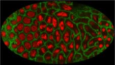
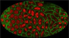
Membrane GFP (Spider-GFP) and with Histone-RFP expressed in the Drosophila embryo. The image is compiled on the Zeiss LSM710 confocal microscope at IISER, Pune by Aparna Sherlekar and Richa Rikhy. The image represents snapshots throughout the syncytial division cycle in the Drosophila embryo at different stages of the cell cycle: interphase, prophase, metaphase, analphase and telophase.
With a similar view in mind, microscopists and microscopy facility managers from across India got together to form a microscopy consortium, along the lines of Euro- Bioimaging (http://www.eurobioimaging.eu/) with colleagues who came from Euro Bioimaging Consortium currently located at EMBL at Heidelberg.
At present, apart from National Center for Biological Science (NCBS) and Institute for Stem Cell Biology and Regenerative Medicine (InStem), this consortium includes – Indian Institute Scientific and Educational Research (IISER) Pune, IISER Kolkata, IISER- Mohali, Tata Institute of Fundamental Research (TIFR), National Institute of Immunology (NII) and Center for Cell and Molecular Biology. The idea will be to have microscopy centers spread all over India, where anyone who wishes to avail of a particular technology to solve a scientific problem can do so. Often, labs or groups are not able to do a particular experiment because of lack of expertise, training or access to facilities and instruments. This is precisely what the Microscopy Consortium aims to minimize. In collaboration with Center for Cellular and Molecular Platforms (CCAMP), the consortium will also provide training in any state of the art technology. The microscopy centers will open their facility for external use, allowing groups across India and the world, access to not just the instruments and facility but also to the expertise that the centers host.
Once formed and functional, this will be a scientific facility for researchers across India. If your institute or lab has a microscopic technology that you feel should be a part of the consortium, you are willing to open the facility for external users and participate in training programs, please contact – krishna/mayor (Krishna@ncbs.res.in, mayor@ncbs.res.in)
Further reading: